 Movie
Movie Controller


+ Open data

- Basic information
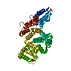
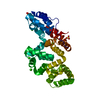
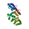
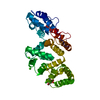
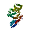
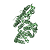
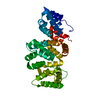
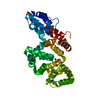
| Entry | Database: PDB / ID: 1n41 | ||||||
|---|---|---|---|---|---|---|---|
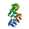
| Title | Crystal Structure of Annexin V K27E Mutant | ||||||
 Components Components | annexin V | ||||||
 Keywords Keywords | LIPID BINDING PROTEIN / calcium / phospholipid membrane binding protein | ||||||
| Function / homology |  Function and homology information Function and homology informationnegative regulation of prolactin secretion / cellular response to gonadotropin-releasing hormone / Platelet degranulation / cellular response to lead ion / regulation of flagellated sperm motility / endothelial microparticle / response to thyroid hormone / P-type calcium transporter activity / calcium-dependent phospholipid binding / vesicle membrane ...negative regulation of prolactin secretion / cellular response to gonadotropin-releasing hormone / Platelet degranulation / cellular response to lead ion / regulation of flagellated sperm motility / endothelial microparticle / response to thyroid hormone / P-type calcium transporter activity / calcium-dependent phospholipid binding / vesicle membrane / phosphatidylserine binding / peptide hormone binding / negative regulation of blood coagulation / intercalated disc / cell projection / axon terminus / sarcolemma / receptor tyrosine kinase binding / response to calcium ion / Z disc / blood coagulation / synaptic vesicle / synaptic vesicle membrane / perikaryon / positive regulation of apoptotic process / external side of plasma membrane / neuronal cell body / calcium ion binding / dendrite / : / identical protein binding / cytoplasm Similarity search - Function | ||||||
| Biological species |  | ||||||
| Method |  X-RAY DIFFRACTION / FOURIER SYNTHESIS / Resolution: 2.1 Å X-RAY DIFFRACTION / FOURIER SYNTHESIS / Resolution: 2.1 Å | ||||||
 Authors Authors | Mo, Y.D. / Campos, B. / Mealy, T.R. / Commodore, L. / Head, J.F. / Dedman, J.R. / Seaton, B.A. | ||||||
 Citation Citation | Journal: J.Biol.Chem. / Year: 2003 Title: Interfacial basic cluster in annexin V couples phospholipid binding and trimer formation on membrane surfaces Authors: Mo, Y.D. / Campos, B. / Mealy, T.R. / Commodore, L. / Head, J.F. / Dedman, J.R. / Seaton, B.A. | ||||||
| History |
|
- Structure visualization
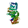
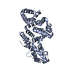
| Structure viewer | Molecule: MolmilJmol/JSmol |
|---|
- Downloads & links
-Download
| PDBx/mmCIF format | 1n41.cif.gz | 80.1 KB | Display | PDBx/mmCIF format |
|---|---|---|---|---|
| PDB format | pdb1n41.ent.gz | 60.2 KB | Display | PDB format |
| PDBx/mmJSON format | 1n41.json.gz | Tree view | PDBx/mmJSON format | |
| Others |  Other downloads Other downloads |
-Validation report
| Arichive directory | https://data.pdbj.org/pub/pdb/validation_reports/n4/1n41ftp://data.pdbj.org/pub/pdb/validation_reports/n4/1n41 | HTTPS FTP |
|---|
-Related structure data


-Links
 PDBj
PDBj- Assembly
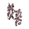
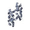
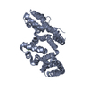
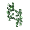
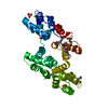
| Deposited unit | 
| ||||||||
|---|---|---|---|---|---|---|---|---|---|
| 1 |
| ||||||||
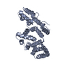
| Unit cell |
|
-Components
| #1: Protein | Mass: 35788.395 Da / Num. of mol.: 1 / Mutation: K27E Source method: isolated from a genetically manipulated source Source: (gene. exp.)  | ||||
|---|---|---|---|---|---|
| #2: Chemical | ChemComp-CA /   Mass: 40.078 Da / Num. of mol.: 5 / Source method: obtained synthetically / Formula: Ca Mass: 40.078 Da / Num. of mol.: 5 / Source method: obtained synthetically / Formula: Ca#3: Chemical |   Mass: 96.063 Da / Num. of mol.: 3 / Source method: obtained synthetically / Formula: SO4 Mass: 96.063 Da / Num. of mol.: 3 / Source method: obtained synthetically / Formula: SO4#4: Water | ChemComp-HOH / |  Mass: 18.015 Da / Num. of mol.: 195 / Source method: isolated from a natural source / Formula: H2O Mass: 18.015 Da / Num. of mol.: 195 / Source method: isolated from a natural source / Formula: H2O |
-Experimental details
-Experiment
| Experiment | Method: X-RAY DIFFRACTION / Number of used crystals: 1 |
|---|
- Sample preparation
| Crystal | Density Matthews: 2.33 Å3/Da / Density % sol: 46.72 % | |||||||||||||||||||||||||||||||||||||||||||||||||
|---|---|---|---|---|---|---|---|---|---|---|---|---|---|---|---|---|---|---|---|---|---|---|---|---|---|---|---|---|---|---|---|---|---|---|---|---|---|---|---|---|---|---|---|---|---|---|---|---|---|---|
| Crystal grow | Temperature: 290 K / Method: vapor diffusion, sitting drop / pH: 8.2 Details: ammonium sulfate, CaCl2, pH 8.2, VAPOR DIFFUSION, SITTING DROP, temperature 290K | |||||||||||||||||||||||||||||||||||||||||||||||||
| Crystal grow | *PLUS Method: vapor diffusion | |||||||||||||||||||||||||||||||||||||||||||||||||
| Components of the solutions | *PLUS
|
-Data collection
| Diffraction | Mean temperature: 293 K |
|---|---|
| Diffraction source | Source: ROTATING ANODE / Type: RIGAKU RU300 / Wavelength: 1.5418 |
| Detector | Type: RIGAKU RAXIS IIC / Detector: IMAGE PLATE |
| Radiation | Protocol: SINGLE WAVELENGTH / Monochromatic (M) / Laue (L): M / Scattering type: x-ray |
| Radiation wavelength | Wavelength: 1.5418 Å / Relative weight: 1 |
| Reflection | Resolution: 2→50 Å / Num. all: 23159 / Num. obs: 21924 / % possible obs: 95 % / Observed criterion σ(F): 0 / Observed criterion σ(I): 0 / Rmerge(I) obs: 0.064 |
| Reflection shell | Resolution: 2→2.07 Å / Rmerge(I) obs: 0.234 / % possible all: 76.4 |
| Reflection | *PLUS Lowest resolution: 50 Å / % possible obs: 96.7 % |
| Reflection shell | *PLUS % possible obs: 92 % / Rmerge(I) obs: 0.236 |
- Processing
| Software |
| ||||||||||||||||||||
|---|---|---|---|---|---|---|---|---|---|---|---|---|---|---|---|---|---|---|---|---|---|
| Refinement | Method to determine structure: FOURIER SYNTHESIS / Resolution: 2.1→50 Å / Cross valid method: THROUGHOUT / σ(F): 0 / Stereochemistry target values: Engh & Huber
| ||||||||||||||||||||
| Refinement step | Cycle: LAST / Resolution: 2.1→50 Å
| ||||||||||||||||||||
| Refine LS restraints |
| ||||||||||||||||||||
| Refinement | *PLUS Lowest resolution: 50 Å / Rfactor obs: 0.187 | ||||||||||||||||||||
| Solvent computation | *PLUS | ||||||||||||||||||||
| Displacement parameters | *PLUS |
