Mass: 18.015 Da / Num. of mol.: 155 / Source method: isolated from a natural source / Formula: H2O
Has protein modification
N
Sequence details
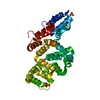
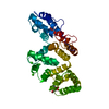
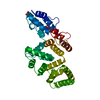
IN THE NATIVE PROTEIN THE N-TERMINAL ALANINE IS ACETYLATED, BUT NO DENSITY IS SEEN FOR THE ...IN THE NATIVE PROTEIN THE N-TERMINAL ALANINE IS ACETYLATED, BUT NO DENSITY IS SEEN FOR THE ACETYLATION, AND THEREFORE HAS BEEN MODELLED AS ALANINE. WHILE IT IS POSSIBLE THAT THE ACETYLATION IS NOT PRESENT IN THE ACTUAL BIOLOGICAL MATERIAL USED, IT IS CERTAINLY REASONABLE TO ASSUME THAT IT IS NOT SEEN IN THE ELECTRON DENSITY MAP.
-
Experimental details
-
Experiment
Experiment
Method: X-RAY DIFFRACTION / Number of used crystals: 1
-
Sample preparation
Crystal
Density Matthews: 2.5 Å3/Da / Density % sol: 50.9 %
Crystal grow
pH: 7.1 Details: PROTEIN CRYSTALLIZED FROM AMMONIUM SULFATE, 20MM CACL2, 50MM HEPES, PH 8.2; SOAKED IN 20% PEG, 20MM CACL2, 50MM GPE, PH 7.1
In the structure databanks used in Yorodumi, some data are registered as the other names, "COVID-19 virus" and "2019-nCoV". Here are the details of the virus and the list of structure data.
Jan 31, 2019. EMDB accession codes are about to change! (news from PDBe EMDB page)
EMDB accession codes are about to change! (news from PDBe EMDB page)
The allocation of 4 digits for EMDB accession codes will soon come to an end. Whilst these codes will remain in use, new EMDB accession codes will include an additional digit and will expand incrementally as the available range of codes is exhausted. The current 4-digit format prefixed with “EMD-” (i.e. EMD-XXXX) will advance to a 5-digit format (i.e. EMD-XXXXX), and so on. It is currently estimated that the 4-digit codes will be depleted around Spring 2019, at which point the 5-digit format will come into force.
The EM Navigator/Yorodumi systems omit the EMD- prefix.
Related info.:Q: What is EMD? / ID/Accession-code notation in Yorodumi/EM Navigator
Yorodumi is a browser for structure data from EMDB, PDB, SASBDB, etc.
This page is also the successor to EM Navigator detail page, and also detail information page/front-end page for Omokage search.
The word "yorodu" (or yorozu) is an old Japanese word meaning "ten thousand". "mi" (miru) is to see.
Related info.:EMDB / PDB / SASBDB / Comparison of 3 databanks / Yorodumi Search / Aug 31, 2016. New EM Navigator & Yorodumi / Yorodumi Papers / Jmol/JSmol / Function and homology information / Changes in new EM Navigator and Yorodumi
 Movie
Movie Controller


 Open data
 Basic information
 Components
 Keywords
 Function and homology information
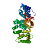
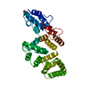
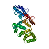
 X-RAY DIFFRACTION / DIFFERENCE FOURIER / Resolution: 1.9 Å
  Authors
 Citation
 Structure visualization
 Downloads & links
 Other downloads


 PDBj
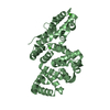
PDBj Assembly

 Mass: 40.078 Da / Num. of mol.: 10 / Source method: obtained synthetically / Formula: Ca
 Mass: 215.142 Da / Num. of mol.: 1 / Source method: obtained synthetically / Formula: C5H14NO6P
 Mass: 18.015 Da / Num. of mol.: 155 / Source method: isolated from a natural source / Formula: H2O
 Sample preparation
 Processing