| 登録情報 | データベース: PDB / ID: 1n3o
|
|---|
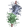
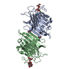
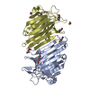
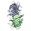
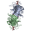
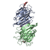
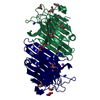
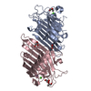
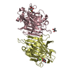
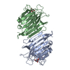
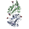
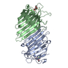
| タイトル | Pterocarcpus angolensis lectin in complex with alpha-methyl glucose |
|---|
 要素 要素 | lectin PAL |
|---|
 キーワード キーワード | SUGAR BINDING PROTEIN / lectin / carbohydrate recognition / glucose |
|---|
| 機能・相同性 |  機能・相同性情報 機能・相同性情報
Legume lectin / Legume lectin, alpha chain, conserved site / Legume lectins alpha-chain signature. / Legume lectins beta-chain signature. / Legume lectin domain / Legume lectin domain / : / Legume lectin, beta chain, Mn/Ca-binding site / Jelly Rolls - #200 / Concanavalin A-like lectin/glucanase domain superfamily ...Legume lectin / Legume lectin, alpha chain, conserved site / Legume lectins alpha-chain signature. / Legume lectins beta-chain signature. / Legume lectin domain / Legume lectin domain / : / Legume lectin, beta chain, Mn/Ca-binding site / Jelly Rolls - #200 / Concanavalin A-like lectin/glucanase domain superfamily / Jelly Rolls / Sandwich / Mainly Beta類似検索 - ドメイン・相同性 |
|---|
| 生物種 |  Pterocarpus angolensis (マメ科) Pterocarpus angolensis (マメ科) |
|---|
| 手法 |  X線回折 / シンクロトロン / 分子置換 / 解像度: 2 Å X線回折 / シンクロトロン / 分子置換 / 解像度: 2 Å |
|---|
 データ登録者 データ登録者 | Loris, R. / Imberty, A. / Beeckmans, S. / Van Driessche, E. / Read, J.S. / Bouckaert, J. / De Greve, H. / Buts, L. / Wyns, L. |
|---|
 引用 引用 | ジャーナル: J.BIOL.CHEM. / 年: 2003
タイトル: Crystal structure of Pterocarpus angolensis lectin in complex with glucose, sucrose, and turanose
著者: Loris, R. / Imberty, A. / Beeckmans, S. / Van Driessche, E. / Read, J.S. / Bouckaert, J. / De Greve, H. / Buts, L. / Wyns, L. |
|---|
| 履歴 | | 登録 | 2002年10月29日 | 登録サイト: RCSB / 処理サイト: PDBJ |
|---|
| 改定 1.0 | 2002年11月20日 | Provider: repository / タイプ: Initial release |
|---|
| 改定 1.1 | 2008年4月28日 | Group: Version format compliance |
|---|
| 改定 1.2 | 2011年7月13日 | Group: Version format compliance |
|---|
| 改定 1.3 | 2020年7月29日 | Group: Data collection / Derived calculations / Structure summary
カテゴリ: chem_comp / diffrn_source ...chem_comp / diffrn_source / entity / pdbx_chem_comp_identifier / pdbx_entity_nonpoly / pdbx_struct_conn_angle / struct_conn / struct_site / struct_site_gen
Item: _chem_comp.mon_nstd_flag / _chem_comp.name ..._chem_comp.mon_nstd_flag / _chem_comp.name / _diffrn_source.pdbx_synchrotron_site / _entity.pdbx_description / _pdbx_entity_nonpoly.name / _pdbx_struct_conn_angle.ptnr1_auth_asym_id / _pdbx_struct_conn_angle.ptnr1_auth_comp_id / _pdbx_struct_conn_angle.ptnr1_auth_seq_id / _pdbx_struct_conn_angle.ptnr1_label_asym_id / _pdbx_struct_conn_angle.ptnr1_label_atom_id / _pdbx_struct_conn_angle.ptnr1_label_comp_id / _pdbx_struct_conn_angle.ptnr1_label_seq_id / _pdbx_struct_conn_angle.ptnr2_auth_asym_id / _pdbx_struct_conn_angle.ptnr2_auth_comp_id / _pdbx_struct_conn_angle.ptnr2_auth_seq_id / _pdbx_struct_conn_angle.ptnr2_label_asym_id / _pdbx_struct_conn_angle.ptnr2_label_atom_id / _pdbx_struct_conn_angle.ptnr2_label_comp_id / _pdbx_struct_conn_angle.ptnr3_auth_asym_id / _pdbx_struct_conn_angle.ptnr3_auth_comp_id / _pdbx_struct_conn_angle.ptnr3_auth_seq_id / _pdbx_struct_conn_angle.ptnr3_label_asym_id / _pdbx_struct_conn_angle.ptnr3_label_atom_id / _pdbx_struct_conn_angle.ptnr3_label_comp_id / _pdbx_struct_conn_angle.ptnr3_label_seq_id / _pdbx_struct_conn_angle.value / _struct_conn.pdbx_dist_value / _struct_conn.ptnr1_auth_asym_id / _struct_conn.ptnr1_auth_comp_id / _struct_conn.ptnr1_auth_seq_id / _struct_conn.ptnr1_label_asym_id / _struct_conn.ptnr1_label_atom_id / _struct_conn.ptnr1_label_comp_id / _struct_conn.ptnr1_label_seq_id / _struct_conn.ptnr2_auth_asym_id / _struct_conn.ptnr2_auth_comp_id / _struct_conn.ptnr2_auth_seq_id / _struct_conn.ptnr2_label_asym_id / _struct_conn.ptnr2_label_atom_id / _struct_conn.ptnr2_label_comp_id / _struct_conn.ptnr2_label_seq_id
解説: Carbohydrate remediation / Provider: repository / タイプ: Remediation |
|---|
| 改定 1.4 | 2024年3月13日 | Group: Data collection / Database references / Structure summary
カテゴリ: chem_comp / chem_comp_atom ...chem_comp / chem_comp_atom / chem_comp_bond / database_2
Item: _chem_comp.pdbx_synonyms / _database_2.pdbx_DOI / _database_2.pdbx_database_accession |
|---|
|
|---|
 ムービー
ムービー コントローラー


 データを開く
 基本情報
基本情報 構造の表示
構造の表示 ダウンロードとリンク
 その他のダウンロード


 PDBj
PDBj

 集合体
集合体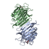

 タイプ: D-saccharide / 分子量: 194.182 Da / 分子数: 2 / 由来タイプ: 組換発現 / 式: C7H14O6
 分子量: 54.938 Da / 分子数: 2 / 由来タイプ: 合成 / 式: Mn
 分子量: 40.078 Da / 分子数: 2 / 由来タイプ: 合成 / 式: Ca
 分子量: 18.015 Da / 分子数: 211 / 由来タイプ: 天然 / 式: H2O
 試料調製
試料調製 / ビームライン: BW7B / 波長: 0.84 Å
 解析
解析