 ムービー
ムービー コントローラー


+ データを開く

- 基本情報
基本情報
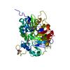
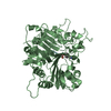
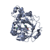
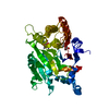
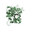
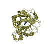
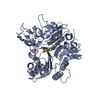
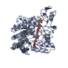
| 登録情報 | データベース: PDB / ID: 1mu7 | ||||||
|---|---|---|---|---|---|---|---|
| タイトル | Crystal Structure of a Human Tyrosyl-DNA Phosphodiesterase (Tdp1)-Tungstate Complex | ||||||
 要素 要素 | Tyrosyl-DNA Phosphodiesterase | ||||||
 キーワード キーワード | HYDROLASE / PLD Superfamily / Protein-Tungstate Complex | ||||||
| 機能・相同性 |  機能・相同性情報 機能・相同性情報3'-tyrosyl-DNA phosphodiesterase activity / single strand break repair / 加水分解酵素; エステル加水分解酵素; リン酸ジエステル加水分解酵素 / exonuclease activity / Nonhomologous End-Joining (NHEJ) / double-strand break repair / single-stranded DNA binding / double-stranded DNA binding / DNA repair / nucleoplasm ...3'-tyrosyl-DNA phosphodiesterase activity / single strand break repair / 加水分解酵素; エステル加水分解酵素; リン酸ジエステル加水分解酵素 / exonuclease activity / Nonhomologous End-Joining (NHEJ) / double-strand break repair / single-stranded DNA binding / double-stranded DNA binding / DNA repair / nucleoplasm / nucleus / plasma membrane / cytoplasm 類似検索 - 分子機能 | ||||||
| 生物種 |  Homo sapiens (ヒト) Homo sapiens (ヒト) | ||||||
| 手法 |  X線回折 / シンクロトロン / 分子置換 / 解像度: 2 Å X線回折 / シンクロトロン / 分子置換 / 解像度: 2 Å | ||||||
 データ登録者 データ登録者 | Davies, D.R. / Interthal, H. / Champoux, J.J. / Hol, W.G.J. | ||||||
 引用 引用 | ジャーナル: J.Mol.Biol. / 年: 2003 タイトル: Insights Into Substrate Binding and Catalytic Mechanism of Human Tyrosyl-DNA Phosphodiesterase (Tdp1) from Vanadate- and Tungstate-Inhibited Structures 著者: Davies, D.R. / Interthal, H. / Champoux, J.J. / Hol, W.G.J. #1: ジャーナル: Structure / 年: 2002タイトル: The crystal structure of human tyrosyl-DNA phosphodiesterase, Tdp1 著者: Davies, D.R. / Interthal, H. / Champoux, J.J. / Hol, W.G.J. | ||||||
| 履歴 |
|
- 構造の表示
構造の表示
| 構造ビューア | 分子: MolmilJmol/JSmol |
|---|
- ダウンロードとリンク
-ダウンロード
| PDBx/mmCIF形式 | 1mu7.cif.gz | 188.6 KB | 表示 | PDBx/mmCIF形式 |
|---|---|---|---|---|
| PDB形式 | pdb1mu7.ent.gz | 147.7 KB | 表示 | PDB形式 |
| PDBx/mmJSON形式 | 1mu7.json.gz | ツリー表示 | PDBx/mmJSON形式 | |
| その他 |  その他のダウンロード その他のダウンロード |
-検証レポート
| アーカイブディレクトリ | https://data.pdbj.org/pub/pdb/validation_reports/mu/1mu7ftp://data.pdbj.org/pub/pdb/validation_reports/mu/1mu7 | HTTPS FTP |
|---|
-関連構造データ


-リンク
 PDBj
PDBj

- 集合体
集合体
| 登録構造単位 | 
| ||||||||
|---|---|---|---|---|---|---|---|---|---|
| 1 | 
| ||||||||
| 2 | 
| ||||||||
| 単位格子 |
| ||||||||
| 詳細 | The biological assembly is a monomer. There are two monomers per asymmetric unit, with the A monomer having the higher occupancy of bound tungstate. |
-要素
| #1: タンパク質 | 分子量: 54731.195 Da / 分子数: 2 / 断片: RESIDUES 149-608 / 変異: D322N,M328T,F548L / 由来タイプ: 組換発現 / 由来: (組換発現) Homo sapiens (ヒト) / プラスミド: pET15b / 生物種 (発現宿主): Escherichia coli / 発現宿主:  #2: 化合物 |   分子量: 247.838 Da / 分子数: 2 / 由来タイプ: 合成 / 式: WO4 分子量: 247.838 Da / 分子数: 2 / 由来タイプ: 合成 / 式: WO4#3: 化合物 |   分子量: 92.094 Da / 分子数: 2 / 由来タイプ: 合成 / 式: C3H8O3 分子量: 92.094 Da / 分子数: 2 / 由来タイプ: 合成 / 式: C3H8O3#4: 水 | ChemComp-HOH / |  分子量: 18.015 Da / 分子数: 307 / 由来タイプ: 天然 / 式: H2O 分子量: 18.015 Da / 分子数: 307 / 由来タイプ: 天然 / 式: H2O |
|---|
-実験情報
-実験
| 実験 | 手法: X線回折 / 使用した結晶の数: 1 |
|---|
- 試料調製
試料調製
| 結晶 | マシュー密度: 2.34 Å3/Da / 溶媒含有率: 47.47 % | |||||||||||||||||||||||||||||||||||||||||||||||||||||||||||||||||||||||||||||
|---|---|---|---|---|---|---|---|---|---|---|---|---|---|---|---|---|---|---|---|---|---|---|---|---|---|---|---|---|---|---|---|---|---|---|---|---|---|---|---|---|---|---|---|---|---|---|---|---|---|---|---|---|---|---|---|---|---|---|---|---|---|---|---|---|---|---|---|---|---|---|---|---|---|---|---|---|---|---|
| 結晶化 | 温度: 298 K / 手法: 蒸気拡散法, シッティングドロップ法 / pH: 7.8 詳細: PEG 8000, sodium chloride, HEPES, spermine, pH 7.8, VAPOR DIFFUSION, SITTING DROP, temperature 298K | |||||||||||||||||||||||||||||||||||||||||||||||||||||||||||||||||||||||||||||
| 結晶化 | *PLUS pH: 8.2 | |||||||||||||||||||||||||||||||||||||||||||||||||||||||||||||||||||||||||||||
| 溶液の組成 | *PLUS
|
-データ収集
| 回折 | 平均測定温度: 113 K |
|---|---|
| 放射光源 | 由来: シンクロトロン / サイト: APS  / ビームライン: 19-ID / 波長: 0.979 Å / ビームライン: 19-ID / 波長: 0.979 Å |
| 検出器 | タイプ: CUSTOM-MADE / 検出器: CCD / 日付: 2002年2月26日 |
| 放射 | プロトコル: SINGLE WAVELENGTH / 単色(M)・ラウエ(L): M / 散乱光タイプ: x-ray |
| 放射波長 | 波長: 0.979 Å / 相対比: 1 |
| 反射 | 解像度: 2→46.3 Å / Num. obs: 74158 / % possible obs: 99 % / Observed criterion σ(F): 0 / Observed criterion σ(I): 0 / 冗長度: 6.58 % / Rsym value: 0.112 / Net I/σ(I): 9.24 |
| 反射 シェル | 解像度: 2→2.07 Å / Mean I/σ(I) obs: 2.18 / Rsym value: 0.409 / % possible all: 92.7 |
| 反射 | *PLUS 最高解像度: 2 Å / Num. obs: 70186 / Rmerge(I) obs: 0.112 |
| 反射 シェル | *PLUS % possible obs: 92.7 % / Rmerge(I) obs: 0.409 |
- 解析
解析
| ソフトウェア |
| ||||||||||||||||||||
|---|---|---|---|---|---|---|---|---|---|---|---|---|---|---|---|---|---|---|---|---|---|
| 精密化 | 構造決定の手法: 分子置換 開始モデル: PDB Entry 1jy1 解像度: 2→46.3 Å / Isotropic thermal model: isotropic / σ(F): 0 / 立体化学のターゲット値: Engh & Huber 詳細: The density is better defined for bound tungstate in the A subunit.
| ||||||||||||||||||||
| 精密化ステップ | サイクル: LAST / 解像度: 2→46.3 Å
| ||||||||||||||||||||
| 拘束条件 |
| ||||||||||||||||||||
| 精密化 | *PLUS 最低解像度: 100 Å / % reflection Rfree: 5 % | ||||||||||||||||||||
| 溶媒の処理 | *PLUS | ||||||||||||||||||||
| 原子変位パラメータ | *PLUS | ||||||||||||||||||||
| 拘束条件 | *PLUS
|
