Mass: 18.015 Da / Num. of mol.: 215 / Source method: isolated from a natural source / Formula: H2O
Has protein modification
N
-
Experimental details
-
Experiment
Experiment
Method: X-RAY DIFFRACTION / Number of used crystals: 1
-
Sample preparation
Crystal
Density Matthews: 2.44 Å3/Da / Density % sol: 49.57 %
Crystal grow
Temperature: 298 K / Method: vapor diffusion, hanging drop / pH: 7.5 Details: The native crystal of yPDE1 (6 mg/ml) was grown by hanging drop against a buffer of 20 mM HEPES pH 7.5, 5% PEG3350, 40 mM Li sulfate, and 12% MPD at room temperature, or a similar buffer but ...Details: The native crystal of yPDE1 (6 mg/ml) was grown by hanging drop against a buffer of 20 mM HEPES pH 7.5, 5% PEG3350, 40 mM Li sulfate, and 12% MPD at room temperature, or a similar buffer but 15% glycerol and 20% MPD, VAPOR DIFFUSION, HANGING DROP, temperature 298K
Method to determine structure: SAD / Resolution: 1.31→34.34 Å / Cor.coef. Fo:Fc: 0.967 / Cor.coef. Fo:Fc free: 0.966 / SU B: 1.215 / SU ML: 0.024 / Cross valid method: THROUGHOUT / ESU R: 0.05 / ESU R Free: 0.044 / Stereochemistry target values: MAXIMUM LIKELIHOOD / Details: HYDROGENS HAVE BEEN USED IF PRESENT IN THE INPUT
Rfactor
Num. reflection
% reflection
Selection details
Rfree
0.17914
4910
5 %
RANDOM
Rwork
0.16806
-
-
-
obs
0.16864
93308
99.52 %
-
all
-
98268
-
-
Solvent computation
Ion probe radii: 0.8 Å / Shrinkage radii: 0.8 Å / VDW probe radii: 1.2 Å / Solvent model: MASK
Displacement parameters
Biso mean: 19.234 Å2
Baniso -1
Baniso -2
Baniso -3
1-
-0.01 Å2
0 Å2
0 Å2
2-
-
0.06 Å2
0 Å2
3-
-
-
-0.06 Å2
Refinement step
Cycle: LAST / Resolution: 1.31→34.34 Å
Protein
Nucleic acid
Ligand
Solvent
Total
Num. atoms
2913
0
34
215
3162
Refine LS restraints
Refine-ID
Type
Dev ideal
Dev ideal target
Number
X-RAY DIFFRACTION
r_bond_refined_d
0.008
0.02
3021
X-RAY DIFFRACTION
r_angle_refined_deg
1.25
1.987
4087
X-RAY DIFFRACTION
r_dihedral_angle_1_deg
6.431
5
361
X-RAY DIFFRACTION
r_dihedral_angle_2_deg
36.912
24.444
135
X-RAY DIFFRACTION
r_dihedral_angle_3_deg
10.344
15
537
X-RAY DIFFRACTION
r_dihedral_angle_4_deg
18.11
15
15
X-RAY DIFFRACTION
r_chiral_restr
0.086
0.2
460
X-RAY DIFFRACTION
r_gen_planes_refined
0.006
0.021
2242
X-RAY DIFFRACTION
r_rigid_bond_restr
2.588
3
3021
X-RAY DIFFRACTION
r_sphericity_free
14.527
5
60
X-RAY DIFFRACTION
r_sphericity_bonded
6.219
5
3103
LS refinement shell
Resolution: 1.31→1.344 Å / Total num. of bins used: 20
Rfactor
Num. reflection
% reflection
Rfree
0.299
317
-
Rwork
0.302
6318
-
obs
-
-
95.58 %
+
About Yorodumi
-
News
-
Feb 9, 2022. New format data for meta-information of EMDB entries
New format data for meta-information of EMDB entries
Version 3 of the EMDB header file is now the official format.
The previous official version 1.9 will be removed from the archive.
In the structure databanks used in Yorodumi, some data are registered as the other names, "COVID-19 virus" and "2019-nCoV". Here are the details of the virus and the list of structure data.
Jan 31, 2019. EMDB accession codes are about to change! (news from PDBe EMDB page)
EMDB accession codes are about to change! (news from PDBe EMDB page)
The allocation of 4 digits for EMDB accession codes will soon come to an end. Whilst these codes will remain in use, new EMDB accession codes will include an additional digit and will expand incrementally as the available range of codes is exhausted. The current 4-digit format prefixed with “EMD-” (i.e. EMD-XXXX) will advance to a 5-digit format (i.e. EMD-XXXXX), and so on. It is currently estimated that the 4-digit codes will be depleted around Spring 2019, at which point the 5-digit format will come into force.
The EM Navigator/Yorodumi systems omit the EMD- prefix.
Related info.:Q: What is EMD? / ID/Accession-code notation in Yorodumi/EM Navigator
Yorodumi is a browser for structure data from EMDB, PDB, SASBDB, etc.
This page is also the successor to EM Navigator detail page, and also detail information page/front-end page for Omokage search.
The word "yorodu" (or yorozu) is an old Japanese word meaning "ten thousand". "mi" (miru) is to see.
Related info.:EMDB / PDB / SASBDB / Comparison of 3 databanks / Yorodumi Search / Aug 31, 2016. New EM Navigator & Yorodumi / Yorodumi Papers / Jmol/JSmol / Function and homology information / Changes in new EM Navigator and Yorodumi
 Movie
Movie Controller

 Yorodumi
 Open data
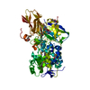
 Basic information
 Components
 Keywords
 Function and homology information
 X-RAY DIFFRACTION /
  Authors
 Citation
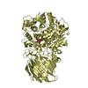
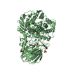
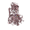
 Structure visualization
 Downloads & links
 Other downloads


 PDBj
PDBj

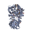
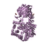
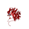
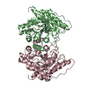
 Assembly


 Mass: 118.174 Da / Num. of mol.: 1 / Source method: obtained synthetically / Formula: C6H14O2 / Comment: precipitant*YM
 Mass: 363.221 Da / Num. of mol.: 1 / Source method: obtained synthetically / Formula: C10H14N5O8P
 Mass: 65.409 Da / Num. of mol.: 2 / Source method: obtained synthetically / Formula: Zn
 Mass: 18.015 Da / Num. of mol.: 215 / Source method: isolated from a natural source / Formula: H2O
 Sample preparation
 / Beamline: X29A / Wavelength: 1.05 Å
 Processing