 Movie
Movie Controller

[English] 日本語
 Yorodumi
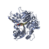
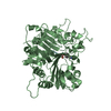
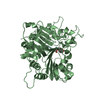
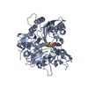
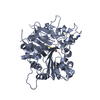
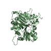
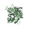
- PDB-1rff: Crystal structure of human Tyrosyl-DNA Phosphodiesterase complexe... -
+ Open data

- Basic information
| Entry | Database: PDB / ID: 1rff | ||||||
|---|---|---|---|---|---|---|---|
| Title | Crystal structure of human Tyrosyl-DNA Phosphodiesterase complexed with vanadate, octapeptide KLNYYDPR, and tetranucleotide AGTT. | ||||||
 Components Components |
| ||||||
 Keywords Keywords | HYDROLASE/DNA / Protein-DNA complex / vanadate complex / transition state mimic / HYDROLASE-DNA COMPLEX | ||||||
| Function / homology |  Function and homology information Function and homology information3'-tyrosyl-DNA phosphodiesterase activity / Hydrolases; Acting on ester bonds; Phosphoric-diester hydrolases / single strand break repair / exonuclease activity / regulation of DNA repair / site of DNA damage / Nonhomologous End-Joining (NHEJ) / double-strand break repair / single-stranded DNA binding / double-stranded DNA binding ...3'-tyrosyl-DNA phosphodiesterase activity / Hydrolases; Acting on ester bonds; Phosphoric-diester hydrolases / single strand break repair / exonuclease activity / regulation of DNA repair / site of DNA damage / Nonhomologous End-Joining (NHEJ) / double-strand break repair / single-stranded DNA binding / double-stranded DNA binding / DNA repair / nucleoplasm / nucleus / cytoplasm Similarity search - Function | ||||||
| Biological species |  Homo sapiens (human) Homo sapiens (human) | ||||||
| Method |  X-RAY DIFFRACTION / SYNCHROTRON / MOLECULAR REPLACEMENT / Resolution: 1.7 Å X-RAY DIFFRACTION / SYNCHROTRON / MOLECULAR REPLACEMENT / Resolution: 1.7 Å | ||||||
 Authors Authors | Davies, D.R. / Interthal, H. / Champoux, J.J. / Hol, W.G. | ||||||
 Citation Citation | Journal: J.Med.Chem. / Year: 2004 Title: Explorations of peptide and oligonucleotide binding sites of tyrosyl-DNA phosphodiesterase using vanadate complexes. Authors: Davies, D.R. / Interthal, H. / Champoux, J.J. / Hol, W.G. #1: Journal: Chem.Biol. / Year: 2003Title: Crystal structure of a transition state mimic for Tdp1 assembled from vanadate, DNA, and a topoisomerase I-derived peptide Authors: Davies, D.R. / Interthal, H. / Champoux, J.J. / Hol, W.G.J. #2: Journal: J.Mol.Biol. / Year: 2002Title: Insights into substrate binding and catalytic mechanism of human Tyrosyl-DNA Phosphodiesterase (Tdp1) from vanadate and tungstate-inhibited structures Authors: Davies, D.R. / Interthal, H. / Champoux, J.J. / Hol, W.G.J. #3: Journal: Structure / Year: 2002Title: The crystal structure of human Tyrosyl-DNA Phosphodiesterase, Tdp1 Authors: Davies, D.R. / Interthal, H. / Champoux, J.J. / Hol, W.G.J. | ||||||
| History |
|
- Structure visualization
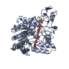
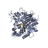
| Structure viewer | Molecule: MolmilJmol/JSmol |
|---|
- Downloads & links
-Download
| PDBx/mmCIF format | 1rff.cif.gz | 199.9 KB | Display | PDBx/mmCIF format |
|---|---|---|---|---|
| PDB format | pdb1rff.ent.gz | 154 KB | Display | PDB format |
| PDBx/mmJSON format | 1rff.json.gz | Tree view | PDBx/mmJSON format | |
| Others |  Other downloads Other downloads |
-Validation report
| Arichive directory | https://data.pdbj.org/pub/pdb/validation_reports/rf/1rffftp://data.pdbj.org/pub/pdb/validation_reports/rf/1rff | HTTPS FTP |
|---|
-Related structure data
| Related structure data |  1rfiC 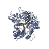 1rg1C  1rg2C  1rgtC  1rguC 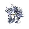 1rh0C 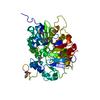 1jy1S S: Starting model for refinement C: citing same article ( |
|---|---|
| Similar structure data |


-Links
 PDBj
PDBj


- Assembly
| Deposited unit | 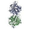
| ||||||||
|---|---|---|---|---|---|---|---|---|---|
| 1 | 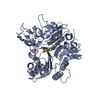
| ||||||||
| 2 | 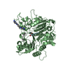
| ||||||||
| Unit cell |
|
-Components
-DNA chain / Protein / Protein/peptide , 3 types, 6 molecules DFABCE
| #1: DNA chain | Mass: 1205.841 Da / Num. of mol.: 2 / Source method: obtained synthetically #2: Protein | Mass: 54731.195 Da / Num. of mol.: 2 / Fragment: residues 149-608 / Mutation: D322N, M328T, F548L Source method: isolated from a genetically manipulated source Source: (gene. exp.) Homo sapiens (human) / Gene: TDP1 / Plasmid: pET15b / Production host:  References: UniProt: Q9NUW8, Hydrolases; Acting on ester bonds; Phosphoric-diester hydrolases #3: Protein/peptide | Mass: 1070.200 Da / Num. of mol.: 2 / Fragment: residues 720-727 / Mutation: L724Y / Source method: obtained synthetically Details: THE Peptide WAS CHEMICALLY SYNTHESIZED. THE SEQUENCE OF THE Peptide IS NATURALLY FOUND IN "Homo sapiens" (Human). Keywords: L724Y |
|---|
-Non-polymers , 3 types, 347 molecules 


| #4: Chemical |  Mass: 114.939 Da / Num. of mol.: 2 / Source method: obtained synthetically / Formula: VO4 Mass: 114.939 Da / Num. of mol.: 2 / Source method: obtained synthetically / Formula: VO4#5: Chemical |  Mass: 202.340 Da / Num. of mol.: 2 / Source method: obtained synthetically / Formula: C10H26N4 Mass: 202.340 Da / Num. of mol.: 2 / Source method: obtained synthetically / Formula: C10H26N4#6: Water | ChemComp-HOH / | Mass: 18.015 Da / Num. of mol.: 343 / Source method: isolated from a natural source / Formula: H2O |
|---|
-Experimental details
-Experiment
| Experiment | Method: X-RAY DIFFRACTION / Number of used crystals: 1 |
|---|
- Sample preparation
| Crystal | Density Matthews: 2.23 Å3/Da / Density % sol: 44.84 % | |||||||||||||||||||||||||||||||||||||||||||||||||||||||||||||||
|---|---|---|---|---|---|---|---|---|---|---|---|---|---|---|---|---|---|---|---|---|---|---|---|---|---|---|---|---|---|---|---|---|---|---|---|---|---|---|---|---|---|---|---|---|---|---|---|---|---|---|---|---|---|---|---|---|---|---|---|---|---|---|---|---|
| Crystal grow | Temperature: 298 K / Method: vapor diffusion, sitting drop / pH: 7.8 Details: PEG 3000, NaCl, HEPES, spermine, pH 7.8, VAPOR DIFFUSION, SITTING DROP, temperature 298K | |||||||||||||||||||||||||||||||||||||||||||||||||||||||||||||||
| Components of the solutions |
| |||||||||||||||||||||||||||||||||||||||||||||||||||||||||||||||
| Crystal grow | *PLUS pH: 8.2 / Method: vapor diffusion, sitting drop | |||||||||||||||||||||||||||||||||||||||||||||||||||||||||||||||
| Components of the solutions | *PLUS
|
-Data collection
| Diffraction | Mean temperature: 100 K |
|---|---|
| Diffraction source | Source: SYNCHROTRON / Site: ALS  / Beamline: 8.2.2 / Wavelength: 1 Å / Beamline: 8.2.2 / Wavelength: 1 Å |
| Detector | Type: ADSC QUANTUM 4 / Detector: CCD / Date: Feb 1, 2003 |
| Radiation | Protocol: SINGLE WAVELENGTH / Monochromatic (M) / Laue (L): M / Scattering type: x-ray |
| Radiation wavelength | Wavelength: 1 Å / Relative weight: 1 |
| Reflection | Resolution: 1.7→50 Å / Num. all: 103225 / Num. obs: 103255 / % possible obs: 90.6 % / Observed criterion σ(F): 0 / Observed criterion σ(I): -3 / Redundancy: 4.52 % / Rsym value: 0.071 / Net I/σ(I): 21.04 |
| Reflection shell | Resolution: 1.7→1.76 Å / Mean I/σ(I) obs: 2 / Rsym value: 0.482 / % possible all: 64.6 |
| Reflection | *PLUS Rmerge(I) obs: 0.071 |
| Reflection shell | *PLUS % possible obs: 64.6 % / Rmerge(I) obs: 0.482 |
- Processing
| Software |
| |||||||||||||||||||||||||||||||||||||||||||||||||||||||||||||||||||||||||||
|---|---|---|---|---|---|---|---|---|---|---|---|---|---|---|---|---|---|---|---|---|---|---|---|---|---|---|---|---|---|---|---|---|---|---|---|---|---|---|---|---|---|---|---|---|---|---|---|---|---|---|---|---|---|---|---|---|---|---|---|---|---|---|---|---|---|---|---|---|---|---|---|---|---|---|---|---|
| Refinement | Method to determine structure: MOLECULAR REPLACEMENT Starting model: PDB entry 1jy1 Resolution: 1.7→50 Å / Cor.coef. Fo:Fc: 0.958 / Cor.coef. Fo:Fc free: 0.951 / SU B: 2.321 / SU ML: 0.074 / Cross valid method: THROUGHOUT / σ(F): 0 / ESU R: 0.114 / ESU R Free: 0.105 / Stereochemistry target values: MAXIMUM LIKELIHOOD
| |||||||||||||||||||||||||||||||||||||||||||||||||||||||||||||||||||||||||||
| Solvent computation | Ion probe radii: 0.8 Å / Shrinkage radii: 0.8 Å / VDW probe radii: 1.4 Å / Solvent model: BABINET MODEL WITH MASK | |||||||||||||||||||||||||||||||||||||||||||||||||||||||||||||||||||||||||||
| Displacement parameters | Biso mean: 24.569 Å2
| |||||||||||||||||||||||||||||||||||||||||||||||||||||||||||||||||||||||||||
| Refinement step | Cycle: LAST / Resolution: 1.7→50 Å
| |||||||||||||||||||||||||||||||||||||||||||||||||||||||||||||||||||||||||||
| Refine LS restraints |
| |||||||||||||||||||||||||||||||||||||||||||||||||||||||||||||||||||||||||||
| LS refinement shell | Resolution: 1.7→1.744 Å / Total num. of bins used: 20 /
| |||||||||||||||||||||||||||||||||||||||||||||||||||||||||||||||||||||||||||
| Refinement | *PLUS Highest resolution: 1.7 Å / Lowest resolution: 50 Å / Rfactor Rfree: 0.213 / Rfactor Rwork: 0.193 | |||||||||||||||||||||||||||||||||||||||||||||||||||||||||||||||||||||||||||
| Solvent computation | *PLUS | |||||||||||||||||||||||||||||||||||||||||||||||||||||||||||||||||||||||||||
| Displacement parameters | *PLUS | |||||||||||||||||||||||||||||||||||||||||||||||||||||||||||||||||||||||||||
| Refine LS restraints | *PLUS
|
