 Movie
Movie Controller

[English] 日本語
 Yorodumi
- PDB-1mu9: Crystal Structure of a Human Tyrosyl-DNA Phosphodiesterase (Tdp1)... -
+ Open data

- Basic information
| Entry | Database: PDB / ID: 1mu9 | ||||||
|---|---|---|---|---|---|---|---|
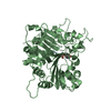
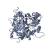
| Title | Crystal Structure of a Human Tyrosyl-DNA Phosphodiesterase (Tdp1)-Vanadate Complex | ||||||
 Components Components | Tyrosyl-DNA Phosphodiesterase | ||||||
 Keywords Keywords | HYDROLASE / PLD Superfamily / Protein-Vanadate Complex | ||||||
| Function / homology |  Function and homology information Function and homology information3'-tyrosyl-DNA phosphodiesterase activity / Hydrolases; Acting on ester bonds; Phosphoric-diester hydrolases / single strand break repair / exonuclease activity / Nonhomologous End-Joining (NHEJ) / double-strand break repair / single-stranded DNA binding / double-stranded DNA binding / DNA repair / nucleoplasm ...3'-tyrosyl-DNA phosphodiesterase activity / Hydrolases; Acting on ester bonds; Phosphoric-diester hydrolases / single strand break repair / exonuclease activity / Nonhomologous End-Joining (NHEJ) / double-strand break repair / single-stranded DNA binding / double-stranded DNA binding / DNA repair / nucleoplasm / nucleus / plasma membrane / cytoplasm Similarity search - Function | ||||||
| Biological species |  Homo sapiens (human) Homo sapiens (human) | ||||||
| Method |  X-RAY DIFFRACTION / SYNCHROTRON / MOLECULAR REPLACEMENT / Resolution: 2.05 Å X-RAY DIFFRACTION / SYNCHROTRON / MOLECULAR REPLACEMENT / Resolution: 2.05 Å | ||||||
 Authors Authors | Davies, D.R. / Interthal, H. / Champoux, J.J. / Hol, W.G.J. | ||||||
 Citation Citation | Journal: J.Mol.Biol. / Year: 2002 Title: Insights Into Substrate Binding and Catalytic Mechanism of Human Tyrosyl-DNA Phosphodiesterase (Tdp1) from Vanadate- and Tungstate-Inhibited Structures Authors: Davies, D.R. / Interthal, H. / Champoux, J.J. / Hol, W.G.J. #1: Journal: Structure / Year: 2002Title: The Crystal Structure of Human Tyrosyl-DNA Phosphodiesterase, Tdp1 Authors: Davies, D.R. / Interthal, H. / Champoux, J.J. / Hol, W.G.J. | ||||||
| History |
|
- Structure visualization
| Structure viewer | Molecule: MolmilJmol/JSmol |
|---|
- Downloads & links
-Download
| PDBx/mmCIF format | 1mu9.cif.gz | 188.4 KB | Display | PDBx/mmCIF format |
|---|---|---|---|---|
| PDB format | pdb1mu9.ent.gz | 147.3 KB | Display | PDB format |
| PDBx/mmJSON format | 1mu9.json.gz | Tree view | PDBx/mmJSON format | |
| Others |  Other downloads Other downloads |
-Validation report
| Arichive directory | https://data.pdbj.org/pub/pdb/validation_reports/mu/1mu9ftp://data.pdbj.org/pub/pdb/validation_reports/mu/1mu9 | HTTPS FTP |
|---|
-Related structure data
| Related structure data |  1mu7C 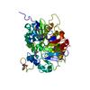 1jy1S S: Starting model for refinement C: citing same article ( |
|---|---|
| Similar structure data |


-Links
 PDBj
PDBj

- Assembly
| Deposited unit | 
| ||||||||
|---|---|---|---|---|---|---|---|---|---|
| 1 | 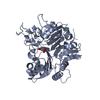
| ||||||||
| 2 | 
| ||||||||
| Unit cell |
| ||||||||
| Details | The biological assembly is a monomer. There are two monomers per asymmetric unit, with the A monomer having the higher occupancy of bound vanadate |
-Components
| #1: Protein | Mass: 54731.195 Da / Num. of mol.: 2 / Fragment: RESIDUES 149-608 / Mutation: D322N,M328T,F548L Source method: isolated from a genetically manipulated source Source: (gene. exp.) Homo sapiens (human) / Plasmid: pET15b / Species (production host): Escherichia coli / Production host:  #2: Chemical |   Mass: 114.939 Da / Num. of mol.: 2 / Source method: obtained synthetically / Formula: VO4 Mass: 114.939 Da / Num. of mol.: 2 / Source method: obtained synthetically / Formula: VO4#3: Chemical |   Mass: 92.094 Da / Num. of mol.: 2 / Source method: obtained synthetically / Formula: C3H8O3 Mass: 92.094 Da / Num. of mol.: 2 / Source method: obtained synthetically / Formula: C3H8O3#4: Water | ChemComp-HOH / |  Mass: 18.015 Da / Num. of mol.: 283 / Source method: isolated from a natural source / Formula: H2O Mass: 18.015 Da / Num. of mol.: 283 / Source method: isolated from a natural source / Formula: H2O |
|---|
-Experimental details
-Experiment
| Experiment | Method: X-RAY DIFFRACTION / Number of used crystals: 1 |
|---|
- Sample preparation
| Crystal | Density Matthews: 2.33 Å3/Da / Density % sol: 47.19 % | |||||||||||||||||||||||||||||||||||||||||||||||||||||||||||||||||||||||||||||
|---|---|---|---|---|---|---|---|---|---|---|---|---|---|---|---|---|---|---|---|---|---|---|---|---|---|---|---|---|---|---|---|---|---|---|---|---|---|---|---|---|---|---|---|---|---|---|---|---|---|---|---|---|---|---|---|---|---|---|---|---|---|---|---|---|---|---|---|---|---|---|---|---|---|---|---|---|---|---|
| Crystal grow | Temperature: 298 K / Method: vapor diffusion, sitting drop / pH: 7.8 Details: PEG 8000, sodium chloride, HEPES, spermine, pH 7.8, VAPOR DIFFUSION, SITTING DROP, temperature 298K | |||||||||||||||||||||||||||||||||||||||||||||||||||||||||||||||||||||||||||||
| Crystal grow | *PLUS pH: 8.2 | |||||||||||||||||||||||||||||||||||||||||||||||||||||||||||||||||||||||||||||
| Components of the solutions | *PLUS
|
-Data collection
| Diffraction | Mean temperature: 113 K |
|---|---|
| Diffraction source | Source: SYNCHROTRON / Site: ALS  / Beamline: 5.0.2 / Wavelength: 1.4 Å / Beamline: 5.0.2 / Wavelength: 1.4 Å |
| Detector | Type: ADSC QUANTUM 4 / Detector: CCD / Date: Mar 20, 2002 |
| Radiation | Protocol: SINGLE WAVELENGTH / Monochromatic (M) / Laue (L): M / Scattering type: x-ray |
| Radiation wavelength | Wavelength: 1.4 Å / Relative weight: 1 |
| Reflection | Resolution: 2.05→19.97 Å / Num. obs: 66910 / % possible obs: 99.8 % / Observed criterion σ(F): 0 / Observed criterion σ(I): 0 / Redundancy: 6.62 % / Rmerge(I) obs: 0.105 / Net I/σ(I): 11.78 |
| Reflection shell | Resolution: 2.05→2.12 Å / Rsym value: 0.387 / % possible all: 100 |
| Reflection | *PLUS Num. obs: 65167 |
| Reflection shell | *PLUS % possible obs: 100 % / Rmerge(I) obs: 0.387 |
- Processing
| Software |
| |||||||||||||||||||||||||
|---|---|---|---|---|---|---|---|---|---|---|---|---|---|---|---|---|---|---|---|---|---|---|---|---|---|---|
| Refinement | Method to determine structure: MOLECULAR REPLACEMENT Starting model: PDB entry 1jy1 Resolution: 2.05→19.97 Å / Isotropic thermal model: isotropic / σ(F): 0 / Stereochemistry target values: Engh & Huber Details: The density is better defined for bound vanadate in the A subunit.
| |||||||||||||||||||||||||
| Refinement step | Cycle: LAST / Resolution: 2.05→19.97 Å
| |||||||||||||||||||||||||
| Refine LS restraints |
| |||||||||||||||||||||||||
| Refinement | *PLUS Lowest resolution: 100 Å / % reflection Rfree: 5 % | |||||||||||||||||||||||||
| Solvent computation | *PLUS | |||||||||||||||||||||||||
| Displacement parameters | *PLUS | |||||||||||||||||||||||||
| Refine LS restraints | *PLUS
|
