 Movie
Movie Controller

[English] 日本語
 Yorodumi
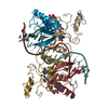
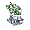
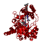
- PDB-1mjo: METHIONINE HOLOREPRESSOR MUTANT (Q44K) PLUS COREPRESSOR (S-ADENOS... -
+ Open data

- Basic information
| Entry | Database: PDB / ID: 1mjo | ||||||
|---|---|---|---|---|---|---|---|
| Title | METHIONINE HOLOREPRESSOR MUTANT (Q44K) PLUS COREPRESSOR (S-ADENOSYL METHIONINE) COMPLEXED TO THE MINIMAL MET CONSENSUS OPERATOR WITH THE CENTRAL TA STEP MUTATED TO AT | ||||||
 Components Components |
| ||||||
 Keywords Keywords | TRANSCRIPTION/DNA / TRANSCRIPTION REGULATION / METJ / METHIONINE REPRESSOR / SHEET-HELIX-HELIX / S-ADENOSYL METHIONINE / DNA / COMPLEX (TRANSCRIPTION REGULATION-DNA) / TRANSCRIPTION-DNA COMPLEX | ||||||
| Function / homology |  Function and homology information Function and homology information: / DNA-binding transcription factor activity / negative regulation of DNA-templated transcription / DNA binding / cytosol Similarity search - Function | ||||||
| Biological species |  | ||||||
| Method |  X-RAY DIFFRACTION / SYNCHROTRON / MOLECULAR REPLACEMENT / Resolution: 2.1 Å X-RAY DIFFRACTION / SYNCHROTRON / MOLECULAR REPLACEMENT / Resolution: 2.1 Å | ||||||
 Authors Authors | Garvie, C.W. / Phillips, S.E.V. | ||||||
 Citation Citation | Journal: Structure Fold.Des. / Year: 2000 Title: Direct and indirect readout in mutant Met repressor-operator complexes. Authors: Garvie, C.W. / Phillips, S.E. | ||||||
| History |
|
- Structure visualization
| Structure viewer | Molecule: MolmilJmol/JSmol |
|---|
- Downloads & links
-Download
| PDBx/mmCIF format | 1mjo.cif.gz | 127.2 KB | Display | PDBx/mmCIF format |
|---|---|---|---|---|
| PDB format | pdb1mjo.ent.gz | 101 KB | Display | PDB format |
| PDBx/mmJSON format | 1mjo.json.gz | Tree view | PDBx/mmJSON format | |
| Others |  Other downloads Other downloads |
-Validation report
| Arichive directory | https://data.pdbj.org/pub/pdb/validation_reports/mj/1mjoftp://data.pdbj.org/pub/pdb/validation_reports/mj/1mjo | HTTPS FTP |
|---|
-Related structure data
| Related structure data |  1mj2C  1mjmC 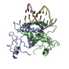 1mjpC  1mjqC  1cmaS C: citing same article ( S: Starting model for refinement |
|---|---|
| Similar structure data |


-Links
 PDBj
PDBj


- Assembly
| Deposited unit | 
| ||||||||
|---|---|---|---|---|---|---|---|---|---|
| 1 |
| ||||||||
| Unit cell |
|
-Components
| #1: DNA chain | Mass: 5819.784 Da / Num. of mol.: 2 / Source method: obtained synthetically #2: Protein | Mass: 12028.607 Da / Num. of mol.: 4 / Mutation: Q44K Source method: isolated from a genetically manipulated source Source: (gene. exp.) #3: Chemical | ChemComp-SAM /   Mass: 398.437 Da / Num. of mol.: 4 / Source method: obtained synthetically / Formula: C15H22N6O5S Mass: 398.437 Da / Num. of mol.: 4 / Source method: obtained synthetically / Formula: C15H22N6O5S#4: Chemical | ChemComp-CA / |   Mass: 40.078 Da / Num. of mol.: 1 / Source method: obtained synthetically / Formula: Ca Mass: 40.078 Da / Num. of mol.: 1 / Source method: obtained synthetically / Formula: Ca#5: Water | ChemComp-HOH / |  Mass: 18.015 Da / Num. of mol.: 510 / Source method: isolated from a natural source / Formula: H2O Mass: 18.015 Da / Num. of mol.: 510 / Source method: isolated from a natural source / Formula: H2O |
|---|
-Experimental details
-Experiment
| Experiment | Method: X-RAY DIFFRACTION / Number of used crystals: 1 |
|---|
- Sample preparation
| Crystal | Density Matthews: 2.92 Å3/Da / Density % sol: 58 % Description: THE FULL METJ COMPLEX WAS USED AS A SEARCH MODEL | ||||||||||||||||||||||||||||||||||||||||||||||||||||||
|---|---|---|---|---|---|---|---|---|---|---|---|---|---|---|---|---|---|---|---|---|---|---|---|---|---|---|---|---|---|---|---|---|---|---|---|---|---|---|---|---|---|---|---|---|---|---|---|---|---|---|---|---|---|---|---|
| Crystal grow | pH: 7 Details: PROTEIN (10MG/ML) + SAM (1MG/ML) + DNA (4MG/ML) WAS CRYSTALLISED FROM 28-38% MPD, 100MM SODIUM CACODYLATE BUFFER, PH 6.0-7.0 | ||||||||||||||||||||||||||||||||||||||||||||||||||||||
| Components of the solutions |
| ||||||||||||||||||||||||||||||||||||||||||||||||||||||
| Crystal | *PLUS | ||||||||||||||||||||||||||||||||||||||||||||||||||||||
| Crystal grow | *PLUS pH: 7 / Method: vapor diffusion, sitting drop | ||||||||||||||||||||||||||||||||||||||||||||||||||||||
| Components of the solutions | *PLUS
|
-Data collection
| Diffraction | Mean temperature: 100 K |
|---|---|
| Diffraction source | Source: SYNCHROTRON / Site: SRS  / Beamline: PX9.5 / Wavelength: 0.92 / Beamline: PX9.5 / Wavelength: 0.92 |
| Detector | Type: MARRESEARCH / Detector: IMAGE PLATE / Date: Jan 1, 1997 / Details: PLATINUM COATED MIRROR |
| Radiation | Monochromator: SILICON (111) / Protocol: SINGLE WAVELENGTH / Monochromatic (M) / Laue (L): M / Scattering type: x-ray |
| Radiation wavelength | Wavelength: 0.92 Å / Relative weight: 1 |
| Reflection | Resolution: 2.1→31.8 Å / Num. obs: 199709 / % possible obs: 99.8 % / Observed criterion σ(I): 3 / Redundancy: 4.7 % / Biso Wilson estimate: 21.5 Å2 / Rmerge(I) obs: 0.044 / Net I/σ(I): 11.3 |
| Reflection shell | Resolution: 2.1→2.17 Å / Redundancy: 4.6 % / Rmerge(I) obs: 0.044 / Mean I/σ(I) obs: 3.8 / Rsym value: 0.201 / % possible all: 100 |
| Reflection | *PLUS Num. obs: 42229 / Num. measured all: 199709 |
| Reflection shell | *PLUS % possible obs: 100 % / Rmerge(I) obs: 0.201 |
- Processing
| Software |
| ||||||||||||||||||||||||||||||||||||||||||||||||||||||||||||
|---|---|---|---|---|---|---|---|---|---|---|---|---|---|---|---|---|---|---|---|---|---|---|---|---|---|---|---|---|---|---|---|---|---|---|---|---|---|---|---|---|---|---|---|---|---|---|---|---|---|---|---|---|---|---|---|---|---|---|---|---|---|
| Refinement | Method to determine structure: MOLECULAR REPLACEMENT Starting model: PDB ENTRY 1CMA Resolution: 2.1→31.8 Å / Rfactor Rfree error: 0.004 / Data cutoff high absF: 10000000 / Data cutoff low absF: 0.001 / Isotropic thermal model: RESTRAINED / Cross valid method: THROUGHOUT / σ(F): 0 / Details: BULK SOLVENT MODEL USED
| ||||||||||||||||||||||||||||||||||||||||||||||||||||||||||||
| Displacement parameters | Biso mean: 35 Å2 | ||||||||||||||||||||||||||||||||||||||||||||||||||||||||||||
| Refine analyze |
| ||||||||||||||||||||||||||||||||||||||||||||||||||||||||||||
| Refinement step | Cycle: LAST / Resolution: 2.1→31.8 Å
| ||||||||||||||||||||||||||||||||||||||||||||||||||||||||||||
| Refine LS restraints |
| ||||||||||||||||||||||||||||||||||||||||||||||||||||||||||||
| LS refinement shell | Resolution: 2.1→2.15 Å / Rfactor Rfree error: 0.021 / Total num. of bins used: 15
| ||||||||||||||||||||||||||||||||||||||||||||||||||||||||||||
| Xplor file |
| ||||||||||||||||||||||||||||||||||||||||||||||||||||||||||||
| Software | *PLUS Name: X-PLOR / Version: 3.86 / Classification: refinement | ||||||||||||||||||||||||||||||||||||||||||||||||||||||||||||
| Refine LS restraints | *PLUS
|
