 Movie
Movie Controller

[English] 日本語
 Yorodumi
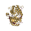
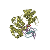
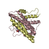
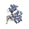
- PDB-1mjp: METHIONINE APOREPRESSOR MUTANT (Q44K) COMPLEXED TO THE MINIMAL ME... -
+ Open data

- Basic information
| Entry | Database: PDB / ID: 1mjp | ||||||
|---|---|---|---|---|---|---|---|
| Title | METHIONINE APOREPRESSOR MUTANT (Q44K) COMPLEXED TO THE MINIMAL MET CONSENSUS OPERATOR | ||||||
 Components Components |
| ||||||
 Keywords Keywords | TRANSCRIPTION/DNA / COMPLEX (TRANSCRIPTION REGULATION-DNA) / METJ / METHIONINE REPRESSOR / SHEET-HELIX-HELIX / SAM / S-ADENOSYL METHIONINE / TRANSCRIPTION-DNA COMPLEX | ||||||
| Function / homology |  Function and homology information Function and homology information: / DNA-binding transcription factor activity / negative regulation of DNA-templated transcription / DNA binding / cytosol Similarity search - Function | ||||||
| Biological species |  | ||||||
| Method |  X-RAY DIFFRACTION / DIFFERENCE FOURIER METHODS / Resolution: 3.4 Å X-RAY DIFFRACTION / DIFFERENCE FOURIER METHODS / Resolution: 3.4 Å | ||||||
 Authors Authors | Garvie, C.W. / Phillips, S.E.V. | ||||||
 Citation Citation | Journal: Structure Fold.Des. / Year: 2000 Title: Direct and indirect readout in mutant Met repressor-operator complexes. Authors: Garvie, C.W. / Phillips, S.E. | ||||||
| History |
|
- Structure visualization
| Structure viewer | Molecule: MolmilJmol/JSmol |
|---|
- Downloads & links
-Download
| PDBx/mmCIF format | 1mjp.cif.gz | 54.7 KB | Display | PDBx/mmCIF format |
|---|---|---|---|---|
| PDB format | pdb1mjp.ent.gz | 43.4 KB | Display | PDB format |
| PDBx/mmJSON format | 1mjp.json.gz | Tree view | PDBx/mmJSON format | |
| Others |  Other downloads Other downloads |
-Validation report
| Arichive directory | https://data.pdbj.org/pub/pdb/validation_reports/mj/1mjpftp://data.pdbj.org/pub/pdb/validation_reports/mj/1mjp | HTTPS FTP |
|---|
-Related structure data
| Related structure data |  1mj2C  1mjmC 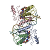 1mjoC  1mjqC  1cmaS C: citing same article ( S: Starting model for refinement |
|---|---|
| Similar structure data |


-Links
 PDBj
PDBj- Assembly
| Deposited unit | 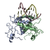
| ||||||||
|---|---|---|---|---|---|---|---|---|---|
| 1 |
| ||||||||
| Unit cell |
|
-Components
| #1: DNA chain | Mass: 3035.003 Da / Num. of mol.: 1 / Source method: obtained synthetically |
|---|---|
| #2: DNA chain | Mass: 2739.823 Da / Num. of mol.: 1 / Source method: obtained synthetically |
| #3: Protein | Mass: 12028.607 Da / Num. of mol.: 2 / Mutation: Q44K Source method: isolated from a genetically manipulated source Source: (gene. exp.) |
-Experimental details
-Experiment
| Experiment | Method: X-RAY DIFFRACTION / Number of used crystals: 1 |
|---|
- Sample preparation
| Crystal | Density Matthews: 2.82 Å3/Da / Density % sol: 56 % | ||||||||||||||||||||||||||||||||||||||||||||||||||||||
|---|---|---|---|---|---|---|---|---|---|---|---|---|---|---|---|---|---|---|---|---|---|---|---|---|---|---|---|---|---|---|---|---|---|---|---|---|---|---|---|---|---|---|---|---|---|---|---|---|---|---|---|---|---|---|---|
| Crystal grow | pH: 7 Details: PROTEIN (10MG/ML) + DNA (4MG/ML) WAS CRYSTALLISED FROM 40% MPD, 100MM SODIUM CACODYLATE BUFFER, PH 7.0. | ||||||||||||||||||||||||||||||||||||||||||||||||||||||
| Components of the solutions |
| ||||||||||||||||||||||||||||||||||||||||||||||||||||||
| Crystal | *PLUS | ||||||||||||||||||||||||||||||||||||||||||||||||||||||
| Crystal grow | *PLUS pH: 7 / Method: vapor diffusion, sitting drop | ||||||||||||||||||||||||||||||||||||||||||||||||||||||
| Components of the solutions | *PLUS
|
-Data collection
| Diffraction | Mean temperature: 100 K |
|---|---|
| Diffraction source | Source: ROTATING ANODE / Type: RIGAKU RU200 / Wavelength: 1.5418 |
| Detector | Type: RIGAKU RAXIS IIC / Detector: IMAGE PLATE / Date: Jun 1, 1997 / Details: MIRRORS |
| Radiation | Protocol: SINGLE WAVELENGTH / Monochromatic (M) / Laue (L): M / Scattering type: x-ray |
| Radiation wavelength | Wavelength: 1.5418 Å / Relative weight: 1 |
| Reflection | Resolution: 3.4→30 Å / Num. obs: 19851 / % possible obs: 99.3 % / Observed criterion σ(I): 3 / Redundancy: 4 % / Biso Wilson estimate: 9.8 Å2 / Rmerge(I) obs: 0.085 / Net I/σ(I): 11.5 |
| Reflection shell | Resolution: 3.4→3.58 Å / Redundancy: 4.1 % / Rmerge(I) obs: 0.085 / Mean I/σ(I) obs: 3.3 / Rsym value: 0.231 / % possible all: 100 |
- Processing
| Software |
| ||||||||||||||||||||||||||||||||||||||||||||||||||||||||||||
|---|---|---|---|---|---|---|---|---|---|---|---|---|---|---|---|---|---|---|---|---|---|---|---|---|---|---|---|---|---|---|---|---|---|---|---|---|---|---|---|---|---|---|---|---|---|---|---|---|---|---|---|---|---|---|---|---|---|---|---|---|---|
| Refinement | Method to determine structure: DIFFERENCE FOURIER METHODS Starting model: PDB ENTRY 1CMA Resolution: 3.4→30 Å / Rfactor Rfree error: 0.014 / Data cutoff high absF: 10000000 / Data cutoff low absF: 0.001 / Isotropic thermal model: GROUP / Cross valid method: THROUGHOUT / σ(F): 0 / Details: BULK SOLVENT MODEL USED
| ||||||||||||||||||||||||||||||||||||||||||||||||||||||||||||
| Displacement parameters | Biso mean: 72.8 Å2
| ||||||||||||||||||||||||||||||||||||||||||||||||||||||||||||
| Refine analyze |
| ||||||||||||||||||||||||||||||||||||||||||||||||||||||||||||
| Refinement step | Cycle: LAST / Resolution: 3.4→30 Å
| ||||||||||||||||||||||||||||||||||||||||||||||||||||||||||||
| Refine LS restraints |
| ||||||||||||||||||||||||||||||||||||||||||||||||||||||||||||
| LS refinement shell | Resolution: 3.4→3.48 Å / Rfactor Rfree error: 0.053 / Total num. of bins used: 15
| ||||||||||||||||||||||||||||||||||||||||||||||||||||||||||||
| Xplor file |
| ||||||||||||||||||||||||||||||||||||||||||||||||||||||||||||
| Software | *PLUS Name: X-PLOR / Version: 3.86 / Classification: refinement | ||||||||||||||||||||||||||||||||||||||||||||||||||||||||||||
| Refine LS restraints | *PLUS
|
