 Movie
Movie Controller

[English] 日本語
 Yorodumi
- PDB-3bw3: Crystal structures and site-directed mutagenesis study of nitroal... -
+ Open data

- Basic information
| Entry | Database: PDB / ID: 3bw3 | ||||||
|---|---|---|---|---|---|---|---|
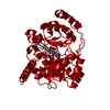
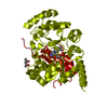
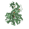
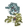
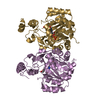
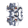
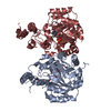
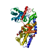
| Title | Crystal structures and site-directed mutagenesis study of nitroalkane oxidase from Streptomyces ansochromogenes | ||||||
 Components Components | 2-nitropropane dioxygenase | ||||||
 Keywords Keywords | OXIDOREDUCTASE / TIM barrel / Dioxygenase | ||||||
| Function / homology |  Function and homology information Function and homology informationnitronate monooxygenase activity / dioxygenase activity / response to toxic substance / nucleotide binding Similarity search - Function | ||||||
| Biological species |  Streptomyces ansochromogenes (bacteria) Streptomyces ansochromogenes (bacteria) | ||||||
| Method |  X-RAY DIFFRACTION / SYNCHROTRON / MOLECULAR REPLACEMENT / Resolution: 2.2 Å X-RAY DIFFRACTION / SYNCHROTRON / MOLECULAR REPLACEMENT / Resolution: 2.2 Å | ||||||
 Authors Authors | Li, Y.H. / Gao, Z.Q. / Hou, H.F. | ||||||
 Citation Citation | Journal: Biochem.Biophys.Res.Commun. / Year: 2011 Title: Crystal structure and site-directed mutagenesis of a nitroalkane oxidase from Streptomyces ansochromogenes Authors: Li, Y.H. / Gao, Z.Q. / Hou, H.F. / Li, L. / Zhang, J.H. / Yang, H.H. / Dong, Y.H. / Tan, H.R. | ||||||
| History |
|
- Structure visualization
| Structure viewer | Molecule: MolmilJmol/JSmol |
|---|
- Downloads & links
-Download
| PDBx/mmCIF format | 3bw3.cif.gz | 82.6 KB | Display | PDBx/mmCIF format |
|---|---|---|---|---|
| PDB format | pdb3bw3.ent.gz | 60.2 KB | Display | PDB format |
| PDBx/mmJSON format | 3bw3.json.gz | Tree view | PDBx/mmJSON format | |
| Others |  Other downloads Other downloads |
-Validation report
| Arichive directory | https://data.pdbj.org/pub/pdb/validation_reports/bw/3bw3ftp://data.pdbj.org/pub/pdb/validation_reports/bw/3bw3 | HTTPS FTP |
|---|
-Related structure data
| Related structure data |  3bw2SC S: Starting model for refinement C: citing same article ( |
|---|---|
| Similar structure data |


-Links
 PDBj
PDBj- Assembly
| Deposited unit | 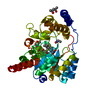
| ||||||||
|---|---|---|---|---|---|---|---|---|---|
| 1 | 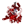
| ||||||||
| Unit cell |
|
-Components
| #1: Protein | Mass: 38521.305 Da / Num. of mol.: 1 / Mutation: H179D Source method: isolated from a genetically manipulated source Source: (gene. exp.) Streptomyces ansochromogenes (bacteria)Strain: 7100 / Gene: 2-npdl / Plasmid: pET23b / Production host: | ||
|---|---|---|---|
| #2: Chemical | ChemComp-FMN /   Mass: 456.344 Da / Num. of mol.: 1 / Source method: obtained synthetically / Formula: C17H21N4O9P Mass: 456.344 Da / Num. of mol.: 1 / Source method: obtained synthetically / Formula: C17H21N4O9P | ||
| #3: Chemical | ChemComp-NIE /   Mass: 75.067 Da / Num. of mol.: 1 / Source method: obtained synthetically / Formula: C2H5NO2 Mass: 75.067 Da / Num. of mol.: 1 / Source method: obtained synthetically / Formula: C2H5NO2 | ||
| #4: Chemical |   Mass: 118.174 Da / Num. of mol.: 2 / Source method: obtained synthetically / Formula: C6H14O2 / Comment: precipitant*YM Mass: 118.174 Da / Num. of mol.: 2 / Source method: obtained synthetically / Formula: C6H14O2 / Comment: precipitant*YM#5: Water | ChemComp-HOH / |  Mass: 18.015 Da / Num. of mol.: 159 / Source method: isolated from a natural source / Formula: H2O Mass: 18.015 Da / Num. of mol.: 159 / Source method: isolated from a natural source / Formula: H2O |
-Experimental details
-Experiment
| Experiment | Method: X-RAY DIFFRACTION / Number of used crystals: 1 |
|---|
- Sample preparation
| Crystal | Density Matthews: 2.41 Å3/Da / Density % sol: 49.05 % |
|---|---|
| Crystal grow | Temperature: 289 K / Method: vapor diffusion, hanging drop / pH: 8.5 Details: 0.2M ammonium dihydrogen phosphate, 0.1M Tris-Cl, 50% MPD, pH8.5, VAPOR DIFFUSION, HANGING DROP, temperature 289K |
-Data collection
| Diffraction | Mean temperature: 100 K |
|---|---|
| Diffraction source | Source: SYNCHROTRON / Site: BSRF  / Beamline: 1W2B / Wavelength: 1 Å / Beamline: 1W2B / Wavelength: 1 Å |
| Detector | Type: MAR scanner 345 mm plate / Detector: IMAGE PLATE / Date: Jun 10, 2007 |
| Radiation | Monochromator: Si 111 CHANNEL / Protocol: SINGLE WAVELENGTH / Monochromatic (M) / Laue (L): M / Scattering type: x-ray |
| Radiation wavelength | Wavelength: 1 Å / Relative weight: 1 |
| Reflection | Resolution: 1.8→10 Å / Num. all: 34891 / Num. obs: 30092 / % possible obs: 86.2 % / Observed criterion σ(F): 1 / Observed criterion σ(I): 1 / Redundancy: 10.5 % / Rmerge(I) obs: 0.08 |
| Reflection shell | Resolution: 1.8→1.86 Å / Redundancy: 10.4 % / Rmerge(I) obs: 0.489 / Num. unique all: 3436 / % possible all: 100 |
- Processing
| Software |
| |||||||||||||||||||||||||
|---|---|---|---|---|---|---|---|---|---|---|---|---|---|---|---|---|---|---|---|---|---|---|---|---|---|---|
| Refinement | Method to determine structure: MOLECULAR REPLACEMENT Starting model: PDB ENTRY 3BW2 Resolution: 2.2→10 Å / σ(F): 1 / Stereochemistry target values: maximum likelihood
| |||||||||||||||||||||||||
| Displacement parameters | Biso mean: 18.67 Å2
| |||||||||||||||||||||||||
| Refinement step | Cycle: LAST / Resolution: 2.2→10 Å
| |||||||||||||||||||||||||
| Refine LS restraints |
| |||||||||||||||||||||||||
| Xplor file |
|
