 Movie
Movie Controller

[English] 日本語
 Yorodumi
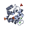
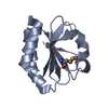
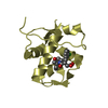
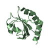
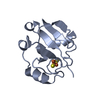
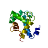
- PDB-1mb0: CRYSTAL STRUCTURE OF THE RESPONSE REGULATOR DIVK AT PH 8.0 IN COM... -
+ Open data

- Basic information
| Entry | Database: PDB / ID: 1mb0 | ||||||
|---|---|---|---|---|---|---|---|
| Title | CRYSTAL STRUCTURE OF THE RESPONSE REGULATOR DIVK AT PH 8.0 IN COMPLEX WITH MN2+ | ||||||
 Components Components | cell division response regulator DivK | ||||||
 Keywords Keywords | SIGNALING PROTEIN / CELL CYCLE / RESPONSE REGULATOR / SIGNAL TRANSDUCTION PROTEIN / Structural Proteomics in Europe / SPINE / Structural Genomics | ||||||
| Function / homology |  Function and homology information Function and homology information | ||||||
| Biological species |  Caulobacter vibrioides (bacteria) Caulobacter vibrioides (bacteria) | ||||||
| Method |  X-RAY DIFFRACTION / SYNCHROTRON / FOURIER SYNTHESIS / Resolution: 2 Å X-RAY DIFFRACTION / SYNCHROTRON / FOURIER SYNTHESIS / Resolution: 2 Å | ||||||
 Authors Authors | Guillet, V. / Ohta, N. / Cabantous, S. / Newton, A. / Samama, J.-P. / Structural Proteomics in Europe (SPINE) | ||||||
 Citation Citation | Journal: J.Biol.Chem. / Year: 2002 Title: Crystallographic and Biochemical Studies of DivK Reveal Novel Features of an Essential Response Regulator in Caulobacter crescentus. Authors: Guillet, V. / Ohta, N. / Cabantous, S. / Newton, A. / Samama, J.-P. #1: Journal: Acta Crystallogr.,Sect.D / Year: 2002Title: Characterization and Crystallization of Divk, an Essential Response Regulator for Cell Division and Differentiation in Caulobacter Crescentus Authors: Cabantous, S. / Guillet, V. / Ohta, N. / Newton, A. / Samama, J.-P. #2: Journal: Embo J. / Year: 1995Title: An Essential Single Domain Response Regulator Required for Normal Cell Division and Differentiation in Caulobacter Crescentus Authors: Hecht, G.B. / Lane, T. / Ohta, N. / Sommer, J.M. / Newton, A. | ||||||
| History |
|
- Structure visualization
| Structure viewer | Molecule: MolmilJmol/JSmol |
|---|
- Downloads & links
-Download
| PDBx/mmCIF format | 1mb0.cif.gz | 36.5 KB | Display | PDBx/mmCIF format |
|---|---|---|---|---|
| PDB format | pdb1mb0.ent.gz | 23.7 KB | Display | PDB format |
| PDBx/mmJSON format | 1mb0.json.gz | Tree view | PDBx/mmJSON format | |
| Others |  Other downloads Other downloads |
-Validation report
| Arichive directory | https://data.pdbj.org/pub/pdb/validation_reports/mb/1mb0ftp://data.pdbj.org/pub/pdb/validation_reports/mb/1mb0 | HTTPS FTP |
|---|
-Related structure data
| Related structure data |  1m5tC 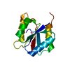 1m5uC 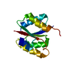 1mavC 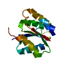 1mb3C C: citing same article ( |
|---|---|
| Similar structure data | |
| Other databases |


-Links
 PDBj
PDBj

- Assembly
| Deposited unit | 
| ||||||||
|---|---|---|---|---|---|---|---|---|---|
| 1 |
| ||||||||
| Unit cell |
|
-Components
| #1: Protein | Mass: 14059.180 Da / Num. of mol.: 1 Source method: isolated from a genetically manipulated source Source: (gene. exp.) Caulobacter vibrioides (bacteria) / Gene: divk / Plasmid details: PT7-7, T7 PROMOTOR / Plasmid: PZHF55 / Production host: | ||
|---|---|---|---|
| #2: Chemical |   Mass: 54.938 Da / Num. of mol.: 2 / Source method: obtained synthetically / Formula: Mn Mass: 54.938 Da / Num. of mol.: 2 / Source method: obtained synthetically / Formula: Mn#3: Water | ChemComp-HOH / |  Mass: 18.015 Da / Num. of mol.: 37 / Source method: isolated from a natural source / Formula: H2O Mass: 18.015 Da / Num. of mol.: 37 / Source method: isolated from a natural source / Formula: H2O |
-Experimental details
-Experiment
| Experiment | Method: X-RAY DIFFRACTION / Number of used crystals: 1 |
|---|
- Sample preparation
| Crystal | Density Matthews: 1.8 Å3/Da / Density % sol: 31 % | ||||||||||||||||||||||||||||||||||||||||||||||||||||||||
|---|---|---|---|---|---|---|---|---|---|---|---|---|---|---|---|---|---|---|---|---|---|---|---|---|---|---|---|---|---|---|---|---|---|---|---|---|---|---|---|---|---|---|---|---|---|---|---|---|---|---|---|---|---|---|---|---|---|
| Crystal grow | Temperature: 285 K / Method: vapor diffusion, hanging or sitting-drop / pH: 8 Details: MES 50mM PH6.0, PEG MME 550 32% AT 285K, The Protein was Concentrated At 2 MG/ML In MES-NAOH PH 6.00 (20 mM), DTT (5 mM) And Mixed With An Equal Volume Of The Reservoir Solution Containing ...Details: MES 50mM PH6.0, PEG MME 550 32% AT 285K, The Protein was Concentrated At 2 MG/ML In MES-NAOH PH 6.00 (20 mM), DTT (5 mM) And Mixed With An Equal Volume Of The Reservoir Solution Containing PEG MME 550 (32%), MES PH 6.00 (40 mM), DTT (5 mM). Crystal Size (300X40X40 microM3) In 20microL Sitting Drops. Crystals Transferred In Reservoir Solutions Whose Ph Was Increased From 6.0 To 8.0 By Steps Of 0.5 Ph Units (The Soaking Time In Each Solution Was 12 Hours). Manganese Derivative Was Obtained By Soaking For 24 Hours Protein Crystals In The Reservoir Solution Supplemented With 10 Mm MnCl2 (PH 8.00). Crystals Were Frozen In Liquid Propane After Soaking For A Few Seconds In Peg MME 550 (50%), Tris PH 8.0 (40 Mm)., VAPOR DIFFUSION, HANGING OR SITTING-DROP | ||||||||||||||||||||||||||||||||||||||||||||||||||||||||
| Crystal grow | *PLUS pH: 6 / Method: vapor diffusion, hanging dropDetails: Cabantous, S., (2002) Acta Crystallogr., Sect.D, 58, 1249. | ||||||||||||||||||||||||||||||||||||||||||||||||||||||||
| Components of the solutions | *PLUS
|
-Data collection
| Diffraction | Mean temperature: 100 K |
|---|---|
| Diffraction source | Source: SYNCHROTRON / Site: ESRF  / Beamline: BM30A / Wavelength: 0.98 / Wavelength: 0.98 Å / Beamline: BM30A / Wavelength: 0.98 / Wavelength: 0.98 Å |
| Detector | Type: MAR scanner 345 mm plate / Detector: IMAGE PLATE / Date: Apr 16, 2000 |
| Radiation | Monochromator: Si111 or Si311 / Protocol: SINGLE WAVELENGTH / Monochromatic (M) / Laue (L): M / Scattering type: x-ray |
| Radiation wavelength | Wavelength: 0.98 Å / Relative weight: 1 |
| Reflection | Resolution: 2→27 Å / Num. obs: 6829 / % possible obs: 98.2 % / Observed criterion σ(I): 0 / Redundancy: 3 % / Biso Wilson estimate: 18.2 Å2 / Rmerge(I) obs: 0.051 / Net I/σ(I): 8.4 |
| Reflection shell | Resolution: 2→2.11 Å / Redundancy: 3.1 % / Rmerge(I) obs: 0.261 / Mean I/σ(I) obs: 2.7 / Num. unique all: 2970 / % possible all: 98.2 |
| Reflection | *PLUS Lowest resolution: 27 Å / Redundancy: 3 % / Num. measured all: 20270 |
| Reflection shell | *PLUS % possible obs: 98.2 % / Mean I/σ(I) obs: 2.7 |
- Processing
| Software |
| ||||||||||||||||||||||||||||||||||||||||||||||||||||||||||||||||||||||||||||||||
|---|---|---|---|---|---|---|---|---|---|---|---|---|---|---|---|---|---|---|---|---|---|---|---|---|---|---|---|---|---|---|---|---|---|---|---|---|---|---|---|---|---|---|---|---|---|---|---|---|---|---|---|---|---|---|---|---|---|---|---|---|---|---|---|---|---|---|---|---|---|---|---|---|---|---|---|---|---|---|---|---|---|
| Refinement | Method to determine structure: FOURIER SYNTHESIS Starting model: APO-DIVK SOLVED AT PH6 Resolution: 2→14.95 Å / Rfactor Rfree error: 0.01 / Isotropic thermal model: RESTRAINED / Cross valid method: THROUGHOUT / σ(F): 0
| ||||||||||||||||||||||||||||||||||||||||||||||||||||||||||||||||||||||||||||||||
| Solvent computation | Solvent model: FLAT MODEL / Bsol: 80.0755 Å2 / ksol: 0.353232 e/Å3 | ||||||||||||||||||||||||||||||||||||||||||||||||||||||||||||||||||||||||||||||||
| Displacement parameters | Biso mean: 32.8 Å2
| ||||||||||||||||||||||||||||||||||||||||||||||||||||||||||||||||||||||||||||||||
| Refine analyze | Luzzati coordinate error free: 0.28 Å / Luzzati sigma a free: 0.26 Å | ||||||||||||||||||||||||||||||||||||||||||||||||||||||||||||||||||||||||||||||||
| Refinement step | Cycle: LAST / Resolution: 2→14.95 Å
| ||||||||||||||||||||||||||||||||||||||||||||||||||||||||||||||||||||||||||||||||
| Refine LS restraints |
| ||||||||||||||||||||||||||||||||||||||||||||||||||||||||||||||||||||||||||||||||
| LS refinement shell | Resolution: 2→2.12 Å / Rfactor Rfree error: 0.028 / Total num. of bins used: 6
| ||||||||||||||||||||||||||||||||||||||||||||||||||||||||||||||||||||||||||||||||
| Xplor file |
| ||||||||||||||||||||||||||||||||||||||||||||||||||||||||||||||||||||||||||||||||
| Refinement | *PLUS Num. reflection obs: 5224 / Rfactor Rfree: 0.243 / Rfactor Rwork: 0.206 | ||||||||||||||||||||||||||||||||||||||||||||||||||||||||||||||||||||||||||||||||
| Solvent computation | *PLUS | ||||||||||||||||||||||||||||||||||||||||||||||||||||||||||||||||||||||||||||||||
| Displacement parameters | *PLUS | ||||||||||||||||||||||||||||||||||||||||||||||||||||||||||||||||||||||||||||||||
| Refine LS restraints | *PLUS
|
