 Movie
Movie Controller


+ Open data

- Basic information
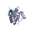
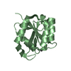
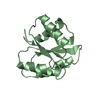
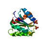
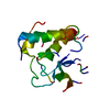
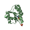
| Entry | Database: PDB / ID: 1m5t | ||||||
|---|---|---|---|---|---|---|---|
| Title | CRYSTAL STRUCTURE OF THE RESPONSE REGULATOR DIVK | ||||||
 Components Components | cell division response regulator DivK | ||||||
 Keywords Keywords | SIGNALING PROTEIN / CELL CYCLE / RESPONSE REGULATOR / SIGNAL TRANSDUCTION PROTEIN / Structural Proteomics in Europe / SPINE / Structural Genomics | ||||||
| Function / homology |  Function and homology information Function and homology information | ||||||
| Biological species |  Caulobacter vibrioides (bacteria) Caulobacter vibrioides (bacteria) | ||||||
| Method |  X-RAY DIFFRACTION / SYNCHROTRON / FOURIER SYNTHESIS / Resolution: 1.6 Å X-RAY DIFFRACTION / SYNCHROTRON / FOURIER SYNTHESIS / Resolution: 1.6 Å | ||||||
 Authors Authors | Guillet, V. / Ohta, N. / Cabantous, S. / Newton, A. / Samama, J.-P. / Structural Proteomics in Europe (SPINE) | ||||||
 Citation Citation | Journal: J.Biol.Chem. / Year: 2002 Title: Crystallographic and biochemical studies of DivK reveal novel features of an essential response regulator in Caulobacter crescentus Authors: Guillet, V. / Ohta, N. / Cabantous, S. / Newton, A. / Samama, J.-P. #1: Journal: Acta Crystallogr.,Sect.D / Year: 2002Title: Characterization and Crystallization of Divk, an Essential Response Regulator for Cell Division and Differentiation in Caulobacter Crescentus Authors: Cabantous, S. / Guillet, V. / Ohta, N. / Newton, A. / Samama, J.-P. #2: Journal: Embo J. / Year: 1995Title: An Essential Single Domain Response Regulator Required for Normal Cell Division and Differentiation in Caulobacter Crescentus Authors: Hecht, G.B. / Lane, T. / Ohta, N. / Sommer, J.M. / Newton, A. | ||||||
| History |
|
- Structure visualization
| Structure viewer | Molecule: MolmilJmol/JSmol |
|---|
- Downloads & links
-Download
| PDBx/mmCIF format | 1m5t.cif.gz | 39.7 KB | Display | PDBx/mmCIF format |
|---|---|---|---|---|
| PDB format | pdb1m5t.ent.gz | 26.8 KB | Display | PDB format |
| PDBx/mmJSON format | 1m5t.json.gz | Tree view | PDBx/mmJSON format | |
| Others |  Other downloads Other downloads |
-Validation report
| Arichive directory | https://data.pdbj.org/pub/pdb/validation_reports/m5/1m5tftp://data.pdbj.org/pub/pdb/validation_reports/m5/1m5t | HTTPS FTP |
|---|
-Related structure data
| Related structure data | 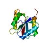 1m5uC 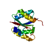 1mavC 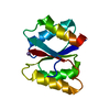 1mb0C 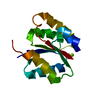 1mb3C C: citing same article ( |
|---|---|
| Similar structure data | |
| Other databases |


-Links
 PDBj
PDBj

- Assembly
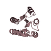
| Deposited unit | 
| ||||||||||
|---|---|---|---|---|---|---|---|---|---|---|---|
| 1 |
| ||||||||||
| Unit cell |
|
-Components
| #1: Protein | Mass: 14059.180 Da / Num. of mol.: 1 Source method: isolated from a genetically manipulated source Details: STRUCTURE AT PH 6.0 IN THE APO-FORM / Source: (gene. exp.) Caulobacter vibrioides (bacteria) / Gene: divk / Plasmid: PT7-7 PZHF55 / Production host: |
|---|---|
| #2: Water | ChemComp-HOH /  Mass: 18.015 Da / Num. of mol.: 120 / Source method: isolated from a natural source / Formula: H2O Mass: 18.015 Da / Num. of mol.: 120 / Source method: isolated from a natural source / Formula: H2O |
-Experimental details
-Experiment
| Experiment | Method: X-RAY DIFFRACTION / Number of used crystals: 1 |
|---|
- Sample preparation
| Crystal | Density Matthews: 1.8 Å3/Da / Density % sol: 31 % | ||||||||||||||||||||||||||||||||||||||||||||||||||||||||
|---|---|---|---|---|---|---|---|---|---|---|---|---|---|---|---|---|---|---|---|---|---|---|---|---|---|---|---|---|---|---|---|---|---|---|---|---|---|---|---|---|---|---|---|---|---|---|---|---|---|---|---|---|---|---|---|---|---|
| Crystal grow | Temperature: 285 K / Method: vapor diffusion, hanging or sitting-drop / pH: 6 Details: MES 50mM PH6.0, PEG MME 550 32% AT 285K, The Protein was Concentrated At 2 MG/ML In MES-NAOH PH 6.00 (20 mM), DTT (5 mM) And Mixed With An Equal Volume Of The Reservoir Solution Containing ...Details: MES 50mM PH6.0, PEG MME 550 32% AT 285K, The Protein was Concentrated At 2 MG/ML In MES-NAOH PH 6.00 (20 mM), DTT (5 mM) And Mixed With An Equal Volume Of The Reservoir Solution Containing PEG MME 550 (32%), MES PH 6.00 (40 mM), DTT (5 mM). Crystal Size (300X40X40 microM3) In 20 microL Sitting Drops. Crystals Were Frozen In Liquid Propane After Soaking For A Few Seconds In PEG MME 550 (50%), MES PH 6.00 (40 mM). VAPOR DIFFUSION, HANGING OR SITTING-DROP at 285K | ||||||||||||||||||||||||||||||||||||||||||||||||||||||||
| Crystal grow | *PLUS pH: 6 / Method: vapor diffusion, hanging dropDetails: Cabantous, S., (2002) Acta Crystallogr., Sect.D, 58, 1249. | ||||||||||||||||||||||||||||||||||||||||||||||||||||||||
| Components of the solutions | *PLUS
|
-Data collection
| Diffraction | Mean temperature: 100 K |
|---|---|
| Diffraction source | Source: SYNCHROTRON / Site: ESRF  / Beamline: ID14-4 / Wavelength: 0.946 / Wavelength: 0.946 Å / Beamline: ID14-4 / Wavelength: 0.946 / Wavelength: 0.946 Å |
| Detector | Type: ADSC QUANTUM 4 / Detector: CCD / Date: Feb 25, 1999 |
| Radiation | Monochromator: Si111 or Si311 crystals / Protocol: SINGLE WAVELENGTH / Monochromatic (M) / Laue (L): M / Scattering type: x-ray |
| Radiation wavelength | Wavelength: 0.946 Å / Relative weight: 1 |
| Reflection | Resolution: 1.6→15 Å / Num. all: 13114 / Num. obs: 13114 / % possible obs: 95.1 % / Observed criterion σ(F): 0 / Observed criterion σ(I): 0 / Redundancy: 4.4 % / Biso Wilson estimate: 14.2 Å2 / Rmerge(I) obs: 0.06 / Net I/σ(I): 6.7 |
| Reflection shell | Resolution: 1.6→1.69 Å / Redundancy: 2.7 % / Rmerge(I) obs: 0.087 / Mean I/σ(I) obs: 5.8 / Num. unique all: 4364 / % possible all: 83 |
| Reflection | *PLUS Highest resolution: 1.6 Å / Lowest resolution: 15 Å / Num. measured all: 58069 / Rmerge(I) obs: 0.06 |
| Reflection shell | *PLUS Highest resolution: 1.6 Å / % possible obs: 83 % / Rmerge(I) obs: 0.087 |
- Processing
| Software |
| ||||||||||||||||||||||||||||||||||||
|---|---|---|---|---|---|---|---|---|---|---|---|---|---|---|---|---|---|---|---|---|---|---|---|---|---|---|---|---|---|---|---|---|---|---|---|---|---|
| Refinement | Method to determine structure: FOURIER SYNTHESIS Starting model: APO-DIVK SOLVED AT PH7 Resolution: 1.6→10 Å / Rfactor Rfree error: 0.006 / Isotropic thermal model: RESTRAINED / Cross valid method: THROUGHOUT / σ(F): 3
| ||||||||||||||||||||||||||||||||||||
| Solvent computation | Solvent model: FLAT MODEL / Bsol: 53.9843 Å2 / ksol: 0.381409 e/Å3 | ||||||||||||||||||||||||||||||||||||
| Displacement parameters | Biso mean: 12 Å2
| ||||||||||||||||||||||||||||||||||||
| Refine analyze | Luzzati coordinate error free: 0.2 Å / Luzzati sigma a free: 0.15 Å | ||||||||||||||||||||||||||||||||||||
| Refinement step | Cycle: LAST / Resolution: 1.6→10 Å
| ||||||||||||||||||||||||||||||||||||
| Refine LS restraints |
| ||||||||||||||||||||||||||||||||||||
| LS refinement shell | Resolution: 1.6→1.7 Å / Rfactor Rfree error: 0.02 / Total num. of bins used: 6
| ||||||||||||||||||||||||||||||||||||
| Xplor file |
| ||||||||||||||||||||||||||||||||||||
| Refinement | *PLUS Highest resolution: 1.6 Å / Lowest resolution: 10 Å / Rfactor obs: 0.193 / Rfactor Rfree: 0.212 / Rfactor Rwork: 0.191 | ||||||||||||||||||||||||||||||||||||
| Solvent computation | *PLUS | ||||||||||||||||||||||||||||||||||||
| Displacement parameters | *PLUS | ||||||||||||||||||||||||||||||||||||
| Refine LS restraints | *PLUS
| ||||||||||||||||||||||||||||||||||||
| LS refinement shell | *PLUS Rfactor Rfree: 0.275 / Rfactor Rwork: 0.21 |
