 Movie
Movie Controller

[English] 日本語
 Yorodumi
- PDB-4f4m: Structure of the type VI peptidoglycan amidase effector Tse1 (C30... -
+ Open data

- Basic information
| Entry | Database: PDB / ID: 4f4m | ||||||
|---|---|---|---|---|---|---|---|
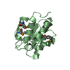
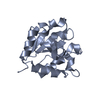
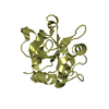
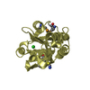
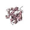
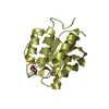
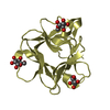
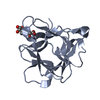
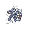
| Title | Structure of the type VI peptidoglycan amidase effector Tse1 (C30A) from Pseudomonas aeruginosa | ||||||
 Components Components | papain peptidoglycan amidase effector Tse1 | ||||||
 Keywords Keywords | Hydrolase regulator / papain peptidoglycan amidase effector / amidase / Tsi1 | ||||||
| Function / homology |  Function and homology information Function and homology informationgamma-D-glutamyl-meso-diaminopimelate peptidase / amidase activity / host cell membrane / extracellular region Similarity search - Function | ||||||
| Biological species |   Pseudomonas aeruginosa (bacteria) Pseudomonas aeruginosa (bacteria) | ||||||
| Method |  X-RAY DIFFRACTION / SYNCHROTRON / MOLECULAR REPLACEMENT / Resolution: 2.677 Å X-RAY DIFFRACTION / SYNCHROTRON / MOLECULAR REPLACEMENT / Resolution: 2.677 Å | ||||||
 Authors Authors | Chou, S. / Mougous, J.D. | ||||||
 Citation Citation | Journal: Cell Rep / Year: 2012 Title: Structure of a peptidoglycan amidase effector targeted to Gram-negative bacteria by the type VI secretion system. Authors: Chou, S. / Bui, N.K. / Russell, A.B. / Lexa, K.W. / Gardiner, T.E. / LeRoux, M. / Vollmer, W. / Mougous, J.D. | ||||||
| History |
|
- Structure visualization
| Structure viewer | Molecule: MolmilJmol/JSmol |
|---|
- Downloads & links
-Download
| PDBx/mmCIF format | 4f4m.cif.gz | 115.3 KB | Display | PDBx/mmCIF format |
|---|---|---|---|---|
| PDB format | pdb4f4m.ent.gz | 90.6 KB | Display | PDB format |
| PDBx/mmJSON format | 4f4m.json.gz | Tree view | PDBx/mmJSON format | |
| Others |  Other downloads Other downloads |
-Validation report
| Arichive directory | https://data.pdbj.org/pub/pdb/validation_reports/f4/4f4mftp://data.pdbj.org/pub/pdb/validation_reports/f4/4f4m | HTTPS FTP |
|---|
-Related structure data


-Links
 PDBj
PDBj- Assembly
| Deposited unit | 
| ||||||||
|---|---|---|---|---|---|---|---|---|---|
| 1 | 
| ||||||||
| 2 | 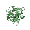
| ||||||||
| 3 | 
| ||||||||
| 4 | 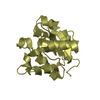
| ||||||||
| Unit cell |
|
-Components
| #1: Protein | Mass: 17255.490 Da / Num. of mol.: 4 / Fragment: Tse1 C30A / Mutation: C30A Source method: isolated from a genetically manipulated source Source: (gene. exp.) Pseudomonas aeruginosa (bacteria) / Gene: PA1844 / Production host: Has protein modification | Y | |
|---|
-Experimental details
-Experiment
| Experiment | Method: X-RAY DIFFRACTION / Number of used crystals: 1 |
|---|
- Sample preparation
| Crystal | Density Matthews: 2.6 Å3/Da / Density % sol: 52.69 % |
|---|---|
| Crystal grow | Temperature: 297 K / Method: vapor diffusion, hanging drop / pH: 7.5 Details: Crystals were obtained by sitting drop vapor diffusion at 25 oC from a 1:1 mixture of 10 mg/mL protein with 0.2 M sodium thiocyanate, 0.1 M HEPES (pH 7.5), 20% PEG3350 for 3 days. Crystals ...Details: Crystals were obtained by sitting drop vapor diffusion at 25 oC from a 1:1 mixture of 10 mg/mL protein with 0.2 M sodium thiocyanate, 0.1 M HEPES (pH 7.5), 20% PEG3350 for 3 days. Crystals were immersed in mother liquor containing 20% glycerol, mounted, and flash frozen in liquid N2. , VAPOR DIFFUSION, HANGING DROP, temperature 297K |
-Data collection
| Diffraction | Mean temperature: 100 K |
|---|---|
| Diffraction source | Source: SYNCHROTRON / Site: ALS  / Beamline: 5.0.2 / Wavelength: 0.979 Å / Beamline: 5.0.2 / Wavelength: 0.979 Å |
| Detector | Type: ADSC QUANTUM 315r / Detector: CCD / Date: Apr 29, 2012 |
| Radiation | Monochromator: Double-crystal, Si(111) / Protocol: SINGLE WAVELENGTH / Monochromatic (M) / Laue (L): M / Scattering type: x-ray |
| Radiation wavelength | Wavelength: 0.979 Å / Relative weight: 1 |
| Reflection | Resolution: 2.677→65.69 Å / Num. all: 19092 / Num. obs: 19072 / % possible obs: 100 % / Biso Wilson estimate: 18.61 Å2 / Rmerge(I) obs: 0.136 |
- Processing
| Software |
| ||||||||||||||||||||||||||||||||||||||||||||||||||||||||
|---|---|---|---|---|---|---|---|---|---|---|---|---|---|---|---|---|---|---|---|---|---|---|---|---|---|---|---|---|---|---|---|---|---|---|---|---|---|---|---|---|---|---|---|---|---|---|---|---|---|---|---|---|---|---|---|---|---|
| Refinement | Method to determine structure: MOLECULAR REPLACEMENT / Resolution: 2.677→65.69 Å / SU ML: 0.31 / σ(F): 1.36 / Phase error: 24.54 / Stereochemistry target values: MLHL
| ||||||||||||||||||||||||||||||||||||||||||||||||||||||||
| Solvent computation | Shrinkage radii: 0.9 Å / VDW probe radii: 1.11 Å / Solvent model: FLAT BULK SOLVENT MODEL | ||||||||||||||||||||||||||||||||||||||||||||||||||||||||
| Refinement step | Cycle: LAST / Resolution: 2.677→65.69 Å
| ||||||||||||||||||||||||||||||||||||||||||||||||||||||||
| Refine LS restraints |
| ||||||||||||||||||||||||||||||||||||||||||||||||||||||||
| LS refinement shell |
|
