 Movie
Movie Controller

[English] 日本語
 Yorodumi
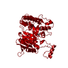
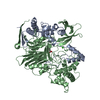
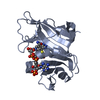
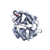
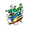
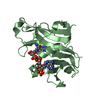
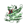
- PDB-1m79: Candida albicans Dihydrofolate Reductase Complexed with Dihydro-N... -
+ Open data

- Basic information
| Entry | Database: PDB / ID: 1m79 | ||||||
|---|---|---|---|---|---|---|---|
| Title | Candida albicans Dihydrofolate Reductase Complexed with Dihydro-Nicotinamide-Adenine-Dinucleotide Phosphate (NADPH) and 5-(4-methoxyphenoxy)-2,4-quinazolinediamine (GW1466) | ||||||
 Components Components | dihydrofolate reductase | ||||||
 Keywords Keywords | OXIDOREDUCTASE / ANTIFUNGAL TARGET / REDUCTASE | ||||||
| Function / homology |  Function and homology information Function and homology informationdihydrofolate metabolic process / dihydrofolate reductase / dihydrofolate reductase activity / folic acid metabolic process / tetrahydrofolate biosynthetic process / one-carbon metabolic process / NADP binding / mitochondrion Similarity search - Function | ||||||
| Biological species |  Candida albicans (yeast) Candida albicans (yeast) | ||||||
| Method |  X-RAY DIFFRACTION / DIRECT REPLACEMENT / Resolution: 1.7 Å X-RAY DIFFRACTION / DIRECT REPLACEMENT / Resolution: 1.7 Å | ||||||
 Authors Authors | Whitlow, M. / Howard, A.J. / Kuyper, L.F. | ||||||
 Citation Citation | Journal: J.Biol.Chem. / Year: 1997 Title: X-Ray Crystallographic Studies of Candida Albicans Dihydrofolate Reductase. High Resolution Structures of the Holoenzyme and an Inhibited Ternary Complex Authors: Whitlow, M. / Howard, A.J. / Stewart, D. / Hardman, K.D. / Kuyper, L.F. / Baccanari, D.P. / Fling, M.E. / Tansik, R.L. #1: Journal: J.Med.Chem. / Year: 2001Title: X-Ray Crystal Structures of Candida Albicans Dihydrofolate Reductase: High Resolution Ternary Complexes in which the Dihydronicotinamide Moiety of Nadph is Displaced by an Inhibitor Authors: Whitlow, M. / Howard, A.J. / Stewart, D. / Hardman, K.D. / Chan, J.H. / Baccanari, D.P. / Tansik, R.L. / Hong, J.S. / Kuyper, L.F. #2: Journal: J.Med.Chem. / Year: 1995Title: Selective Inhibitors of Candida Albicans Dihydrofolate Reductase: Activity and Selectivity of 5-(Arylthio)-2,4-Diaminoquinazolines Authors: Chan, J.H. / Hong, J.S. / Kuyper, L.F. / Baccanari, D.P. / Joyner, S.S. / Tansik, R.L. / Boytos, C.M. / Rudolph, S.K. #3: Journal: J.Biol.Chem. / Year: 1989Title: Characterization of Candida Albicans Dihydrofolate Reductase Authors: Baccanari, D.P. / Tansik, R.L. / Joyner, S.S. / Fling, M.E. / Smith, P.L. / Freisheim, J.H. | ||||||
| History |
|
- Structure visualization
| Structure viewer | Molecule: MolmilJmol/JSmol |
|---|
- Downloads & links
-Download
| PDBx/mmCIF format | 1m79.cif.gz | 106.3 KB | Display | PDBx/mmCIF format |
|---|---|---|---|---|
| PDB format | pdb1m79.ent.gz | 81.6 KB | Display | PDB format |
| PDBx/mmJSON format | 1m79.json.gz | Tree view | PDBx/mmJSON format | |
| Others |  Other downloads Other downloads |
-Validation report
| Arichive directory | https://data.pdbj.org/pub/pdb/validation_reports/m7/1m79ftp://data.pdbj.org/pub/pdb/validation_reports/m7/1m79 | HTTPS FTP |
|---|
-Related structure data
| Related structure data |  1ai9SC 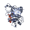 1aoeC 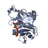 1m78C  1m7aC S: Starting model for refinement C: citing same article ( |
|---|---|
| Similar structure data |


-Links
 PDBj
PDBj

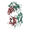
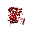
- Assembly
| Deposited unit | 
| ||||||||
|---|---|---|---|---|---|---|---|---|---|
| 1 | 
| ||||||||
| 2 | 
| ||||||||
| Unit cell |
| ||||||||
| Details | Dihydrofolate reductase is active as a monomer. |
-Components
| #1: Protein | Mass: 22194.527 Da / Num. of mol.: 2 Source method: isolated from a genetically manipulated source Source: (gene. exp.) Candida albicans (yeast) / Gene: DFR1 / Plasmid: p1869 / Species (production host): Escherichia coli / Production host:  #2: Chemical |   Mass: 745.421 Da / Num. of mol.: 2 / Source method: obtained synthetically / Formula: C21H30N7O17P3 Mass: 745.421 Da / Num. of mol.: 2 / Source method: obtained synthetically / Formula: C21H30N7O17P3#3: Chemical | 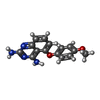  Mass: 282.297 Da / Num. of mol.: 2 / Source method: obtained synthetically / Formula: C15H14N4O2 Mass: 282.297 Da / Num. of mol.: 2 / Source method: obtained synthetically / Formula: C15H14N4O2#4: Chemical |   Mass: 195.237 Da / Num. of mol.: 2 / Source method: obtained synthetically / Formula: C6H13NO4S / Comment: pH buffer*YM Mass: 195.237 Da / Num. of mol.: 2 / Source method: obtained synthetically / Formula: C6H13NO4S / Comment: pH buffer*YM#5: Water | ChemComp-HOH / |  Mass: 18.015 Da / Num. of mol.: 354 / Source method: isolated from a natural source / Formula: H2O Mass: 18.015 Da / Num. of mol.: 354 / Source method: isolated from a natural source / Formula: H2O |
|---|
-Experimental details
-Experiment
| Experiment | Method: X-RAY DIFFRACTION / Number of used crystals: 1 |
|---|
- Sample preparation
| Crystal | Density Matthews: 2.27 Å3/Da / Density % sol: 43 % |
|---|---|
| Crystal grow | Temperature: 277 K / Method: vapor diffusion, hanging drop / pH: 6.5 Details: dihydro-nicotinamide-adenine-dinucleotide phosphate (NADPH), 5-(4-METHOXYPHENOXY)-2,4-QUINAZOLINEDIAMINE (GW1466), PEG-3350, Potassium 4-morphilineethanesulfonic acid (MES), dithiothreitol ...Details: dihydro-nicotinamide-adenine-dinucleotide phosphate (NADPH), 5-(4-METHOXYPHENOXY)-2,4-QUINAZOLINEDIAMINE (GW1466), PEG-3350, Potassium 4-morphilineethanesulfonic acid (MES), dithiothreitol (DTT), pH 6.50, VAPOR DIFFUSION, HANGING DROP, temperature 277K |
-Data collection
| Diffraction | Mean temperature: 293 K |
|---|---|
| Diffraction source | Source: ROTATING ANODE / Type: ELLIOTT GX-21 / Wavelength: 1.5418 / Wavelength: 1.5418 Å |
| Detector | Type: XENTRONICS / Detector: AREA DETECTOR / Date: Mar 16, 1989 / Details: MONOCHROMATOR |
| Radiation | Monochromator: HUBER GRAPHITE MONOCHROMATOR / Protocol: SINGLE WAVELENGTH / Monochromatic (M) / Laue (L): M / Scattering type: x-ray |
| Radiation wavelength | Wavelength: 1.5418 Å / Relative weight: 1 |
| Reflection | Resolution: 1.7→50 Å / Num. obs: 36008 / % possible obs: 83.45 % / Observed criterion σ(F): 0 / Observed criterion σ(I): -3 / Redundancy: 2.45 % / Biso Wilson estimate: 33.74 Å2 / Rmerge(I) obs: 0.0644 / Rsym value: 0.0644 / Net I/σ(I): 21.1 |
| Reflection shell | Resolution: 1.7→1.8 Å / Redundancy: 1.79 % / Rmerge(I) obs: 0.1978 / Mean I/σ(I) obs: 3.7 / Num. unique all: 2562 / Rsym value: 0.1978 / % possible all: 99.9 |
- Processing
| Software |
| ||||||||||||||||||||||||||||||||||||||||||||||||||||||||||||||||||||||||||||
|---|---|---|---|---|---|---|---|---|---|---|---|---|---|---|---|---|---|---|---|---|---|---|---|---|---|---|---|---|---|---|---|---|---|---|---|---|---|---|---|---|---|---|---|---|---|---|---|---|---|---|---|---|---|---|---|---|---|---|---|---|---|---|---|---|---|---|---|---|---|---|---|---|---|---|---|---|---|
| Refinement | Method to determine structure: DIRECT REPLACEMENT Starting model: CANDIDA ALBICANS DHFR (PDB ENTRY 1AI9) Resolution: 1.7→10 Å Isotropic thermal model: Konnert, J.H. & Hendrickson, W.A. (1980) Acta Crystallogr. A A34, 344. σ(F): 2 / σ(I): 0 Stereochemistry target values: Hendrickson, W.A. (1985) Methods Enzymol. 115, 252-270. Details: RESTRAIN LEAST-SQUARES PROCEDURE.
| ||||||||||||||||||||||||||||||||||||||||||||||||||||||||||||||||||||||||||||
| Refinement step | Cycle: LAST / Resolution: 1.7→10 Å
| ||||||||||||||||||||||||||||||||||||||||||||||||||||||||||||||||||||||||||||
| Refine LS restraints |
| ||||||||||||||||||||||||||||||||||||||||||||||||||||||||||||||||||||||||||||
| LS refinement shell |
|
