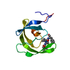
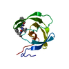
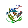
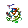
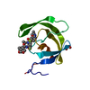
- PDB-1m4c: Crystal Structure of Human Interleukin-2 -
+
Open data
ID or keywords:
Loading...
-
Basic information
Entry
Database: PDB / ID: 1m4c
Title
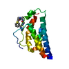
Crystal Structure of Human Interleukin-2
Components
interleukin-2
Keywords
CYTOKINE / four-helix bundle
Function / homology
Function and homology information
response to tacrolimus / kappa-type opioid receptor binding / regulation of CD4-positive, alpha-beta T cell proliferation / regulation of T cell homeostatic proliferation / interleukin-2 receptor binding / negative regulation of lymphocyte proliferation / glycosphingolipid binding / positive regulation of plasma cell differentiation / positive regulation of tissue remodeling / negative regulation of T-helper 17 cell differentiation ...response to tacrolimus / kappa-type opioid receptor binding / regulation of CD4-positive, alpha-beta T cell proliferation / regulation of T cell homeostatic proliferation / interleukin-2 receptor binding / negative regulation of lymphocyte proliferation / glycosphingolipid binding / positive regulation of plasma cell differentiation / positive regulation of tissue remodeling / negative regulation of T-helper 17 cell differentiation / RUNX1 and FOXP3 control the development of regulatory T lymphocytes (Tregs) / leukocyte activation involved in immune response / positive regulation of isotype switching to IgG isotypes / activated T cell proliferation / Interleukin-2 signaling / cell surface receptor signaling pathway via STAT / natural killer cell activation / kinase activator activity / positive regulation of activated T cell proliferation / positive regulation of regulatory T cell differentiation / negative regulation of B cell apoptotic process / interleukin-2-mediated signaling pathway / positive regulation of immunoglobulin production / positive regulation of interleukin-17 production / positive regulation of dendritic spine development / T cell differentiation / Interleukin receptor SHC signaling / extrinsic apoptotic signaling pathway in absence of ligand / positive regulation of B cell proliferation / cytokine activity / growth factor activity / negative regulation of inflammatory response / positive regulation of type II interferon production / positive regulation of inflammatory response / cell-cell signaling / carbohydrate binding / positive regulation of cytosolic calcium ion concentration / RAF/MAP kinase cascade / positive regulation of cell growth / transcription by RNA polymerase II / phospholipase C-activating G protein-coupled receptor signaling pathway / adaptive immune response / response to ethanol / cell adhesion / immune response / positive regulation of cell population proliferation / negative regulation of apoptotic process / positive regulation of transcription by RNA polymerase II / : / extracellular region Similarity search - Function
In the structure databanks used in Yorodumi, some data are registered as the other names, "COVID-19 virus" and "2019-nCoV". Here are the details of the virus and the list of structure data.
Jan 31, 2019. EMDB accession codes are about to change! (news from PDBe EMDB page)
EMDB accession codes are about to change! (news from PDBe EMDB page)
The allocation of 4 digits for EMDB accession codes will soon come to an end. Whilst these codes will remain in use, new EMDB accession codes will include an additional digit and will expand incrementally as the available range of codes is exhausted. The current 4-digit format prefixed with “EMD-” (i.e. EMD-XXXX) will advance to a 5-digit format (i.e. EMD-XXXXX), and so on. It is currently estimated that the 4-digit codes will be depleted around Spring 2019, at which point the 5-digit format will come into force.
The EM Navigator/Yorodumi systems omit the EMD- prefix.
Related info.:Q: What is EMD? / ID/Accession-code notation in Yorodumi/EM Navigator
Yorodumi is a browser for structure data from EMDB, PDB, SASBDB, etc.
This page is also the successor to EM Navigator detail page, and also detail information page/front-end page for Omokage search.
The word "yorodu" (or yorozu) is an old Japanese word meaning "ten thousand". "mi" (miru) is to see.
Related info.:EMDB / PDB / SASBDB / Comparison of 3 databanks / Yorodumi Search / Aug 31, 2016. New EM Navigator & Yorodumi / Yorodumi Papers / Jmol/JSmol / Function and homology information / Changes in new EM Navigator and Yorodumi
 Movie
Movie Controller


 Open data
 Basic information
 Components
 Keywords
 Function and homology information
 Homo sapiens (human)
 X-RAY DIFFRACTION /
  Authors
 Citation
 Structure visualization
 Downloads & links
 Other downloads


 PDBj
PDBj


 Assembly

 Sample preparation
 Processing