 Movie
Movie Controller

[English] 日本語
 Yorodumi
- PDB-1m2w: Pseudomonas fluorescens mannitol 2-dehydrogenase ternary complex ... -
+ Open data

- Basic information
| Entry | Database: PDB / ID: 1m2w | ||||||
|---|---|---|---|---|---|---|---|
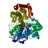
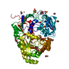
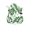
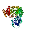
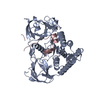
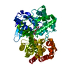
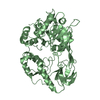
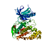
| Title | Pseudomonas fluorescens mannitol 2-dehydrogenase ternary complex with NAD and D-mannitol | ||||||
 Components Components | mannitol dehydrogenase | ||||||
 Keywords Keywords | OXIDOREDUCTASE / Rossmann fold / di-nucleotide binding motif / long-chain dehydrogenase / polyol dehydrogenase / secondary alcohol dehydrogenase | ||||||
| Function / homology |  Function and homology information Function and homology informationmannitol 2-dehydrogenase / mannitol 2-dehydrogenase activity / mannitol metabolic process / nucleotide binding Similarity search - Function | ||||||
| Biological species |  Pseudomonas fluorescens (bacteria) Pseudomonas fluorescens (bacteria) | ||||||
| Method |  X-RAY DIFFRACTION / SYNCHROTRON / MOLECULAR REPLACEMENT / Resolution: 1.8 Å X-RAY DIFFRACTION / SYNCHROTRON / MOLECULAR REPLACEMENT / Resolution: 1.8 Å | ||||||
 Authors Authors | Kavanagh, K.L. / Klimacek, M. / Nidetzky, B. / Wilson, D.K. | ||||||
 Citation Citation | Journal: J.Biol.Chem. / Year: 2002 Title: Crystal Structure of Pseudomonas fluorescens Mannitol 2-Dehydrogenase Binary and Ternary Complexes. Specificity and Catalytic Mechanism Authors: Kavanagh, K.L. / Klimacek, M. / Nidetzky, B. / Wilson, D.K. | ||||||
| History |
|
- Structure visualization
| Structure viewer | Molecule: MolmilJmol/JSmol |
|---|
- Downloads & links
-Download
| PDBx/mmCIF format | 1m2w.cif.gz | 216.9 KB | Display | PDBx/mmCIF format |
|---|---|---|---|---|
| PDB format | pdb1m2w.ent.gz | 173.6 KB | Display | PDB format |
| PDBx/mmJSON format | 1m2w.json.gz | Tree view | PDBx/mmJSON format | |
| Others |  Other downloads Other downloads |
-Validation report
| Arichive directory | https://data.pdbj.org/pub/pdb/validation_reports/m2/1m2wftp://data.pdbj.org/pub/pdb/validation_reports/m2/1m2w | HTTPS FTP |
|---|
-Related structure data
| Related structure data |  1lj8SC S: Starting model for refinement C: citing same article ( |
|---|---|
| Similar structure data |


-Links
 PDBj
PDBj


- Assembly
| Deposited unit | 
| ||||||||||
|---|---|---|---|---|---|---|---|---|---|---|---|
| 1 | 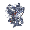
| ||||||||||
| 2 | 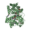
| ||||||||||
| Unit cell |
| ||||||||||
| Components on special symmetry positions |
|
-Components
| #1: Protein | Mass: 55072.531 Da / Num. of mol.: 2 Source method: isolated from a genetically manipulated source Source: (gene. exp.) Pseudomonas fluorescens (bacteria) / Strain: DSM50106 / Gene: mtlD / Production host: #2: Chemical |   Mass: 663.425 Da / Num. of mol.: 2 / Source method: obtained synthetically / Formula: C21H27N7O14P2 / Comment: NAD*YM Mass: 663.425 Da / Num. of mol.: 2 / Source method: obtained synthetically / Formula: C21H27N7O14P2 / Comment: NAD*YM#3: Chemical |   Mass: 182.172 Da / Num. of mol.: 2 / Source method: obtained synthetically / Formula: C6H14O6 / Comment: medication*YM Mass: 182.172 Da / Num. of mol.: 2 / Source method: obtained synthetically / Formula: C6H14O6 / Comment: medication*YM#4: Water | ChemComp-HOH / |  Mass: 18.015 Da / Num. of mol.: 630 / Source method: isolated from a natural source / Formula: H2O Mass: 18.015 Da / Num. of mol.: 630 / Source method: isolated from a natural source / Formula: H2OHas protein modification | Y | |
|---|
-Experimental details
-Experiment
| Experiment | Method: X-RAY DIFFRACTION / Number of used crystals: 1 |
|---|
- Sample preparation
| Crystal | Density Matthews: 2.57 Å3/Da / Density % sol: 52.22 % | ||||||||||||||||||||||||||||||||||||||||||||||||||||||||
|---|---|---|---|---|---|---|---|---|---|---|---|---|---|---|---|---|---|---|---|---|---|---|---|---|---|---|---|---|---|---|---|---|---|---|---|---|---|---|---|---|---|---|---|---|---|---|---|---|---|---|---|---|---|---|---|---|---|
| Crystal grow | Temperature: 293 K / Method: vapor diffusion, hanging drop / pH: 5 Details: 34% PEG 4000, 250 mM ammonium acetate, 100 mM sodium citrate, 10 mM DTT, pH 5.0, VAPOR DIFFUSION, HANGING DROP at 293K | ||||||||||||||||||||||||||||||||||||||||||||||||||||||||
| Crystal grow | *PLUS pH: 7.5 | ||||||||||||||||||||||||||||||||||||||||||||||||||||||||
| Components of the solutions | *PLUS
|
-Data collection
| Diffraction | Mean temperature: 100 K |
|---|---|
| Diffraction source | Source: SYNCHROTRON / Site: SSRL  / Beamline: BL9-1 / Wavelength: 0.9198 Å / Beamline: BL9-1 / Wavelength: 0.9198 Å |
| Detector | Type: MARRESEARCH / Detector: IMAGE PLATE / Date: Apr 24, 2002 |
| Radiation | Monochromator: silicon crystal / Protocol: SINGLE WAVELENGTH / Monochromatic (M) / Laue (L): M / Scattering type: x-ray |
| Radiation wavelength | Wavelength: 0.9198 Å / Relative weight: 1 |
| Reflection | Resolution: 1.8→30 Å / Num. all: 105733 / Num. obs: 104464 / % possible obs: 99.5 % / Observed criterion σ(I): 2 / Redundancy: 3.5 % / Rmerge(I) obs: 0.064 / Net I/σ(I): 18.4 |
| Reflection shell | Resolution: 1.8→1.84 Å / Rmerge(I) obs: 0.371 / Mean I/σ(I) obs: 2.8 / % possible all: 96.1 |
| Reflection | *PLUS Highest resolution: 1.8 Å / Num. obs: 105300 / Num. measured all: 367595 / Rmerge(I) obs: 0.064 |
| Reflection shell | *PLUS Highest resolution: 1.8 Å / % possible obs: 96.1 % / Rmerge(I) obs: 0.371 |
- Processing
| Software |
| |||||||||||||||||||||||||
|---|---|---|---|---|---|---|---|---|---|---|---|---|---|---|---|---|---|---|---|---|---|---|---|---|---|---|
| Refinement | Method to determine structure: MOLECULAR REPLACEMENT Starting model: 1LJ8 Resolution: 1.8→30 Å / Cross valid method: THROUGHOUT / σ(F): 0 / Stereochemistry target values: Engh & Huber
| |||||||||||||||||||||||||
| Refinement step | Cycle: LAST / Resolution: 1.8→30 Å
| |||||||||||||||||||||||||
| Refine LS restraints |
| |||||||||||||||||||||||||
| LS refinement shell | Resolution: 1.8→1.84 Å /
| |||||||||||||||||||||||||
| Refinement | *PLUS Highest resolution: 1.8 Å / Lowest resolution: 30 Å / Rfactor obs: 0.1761 / Rfactor Rfree: 0.202 / Rfactor Rwork: 0.176 | |||||||||||||||||||||||||
| Solvent computation | *PLUS | |||||||||||||||||||||||||
| Displacement parameters | *PLUS |
