 Movie
Movie Controller

[English] 日本語
 Yorodumi
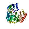
- PDB-6hlx: Structure of the PBP AgaA in complex with agropinic acid from A.t... -
+ Open data

- Basic information
| Entry | Database: PDB / ID: 6hlx | ||||||
|---|---|---|---|---|---|---|---|
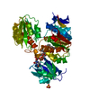
| Title | Structure of the PBP AgaA in complex with agropinic acid from A.tumefacien R10 | ||||||
 Components Components | AgaA | ||||||
 Keywords Keywords | TRANSPORT PROTEIN / periplasmic binding protein / ABC transporter | ||||||
| Function / homology |  Function and homology information Function and homology informationpeptide transport / peptide transmembrane transporter activity / ATP-binding cassette (ABC) transporter complex / outer membrane-bounded periplasmic space / metal ion binding Similarity search - Function | ||||||
| Biological species |  Rhizobium radiobacter (Agrobacterium genomosp. 4) Rhizobium radiobacter (Agrobacterium genomosp. 4) | ||||||
| Method |  X-RAY DIFFRACTION / SYNCHROTRON / MOLECULAR REPLACEMENT / Resolution: 1.65 Å X-RAY DIFFRACTION / SYNCHROTRON / MOLECULAR REPLACEMENT / Resolution: 1.65 Å | ||||||
 Authors Authors | Morera, S. / Marty, L. / Vigouroux, A. | ||||||
 Citation Citation | Journal: Biochem. J. / Year: 2019 Title: Structural basis for two efficient modes of agropinic acid opine import into the bacterial pathogenAgrobacterium tumefaciens. Authors: Marty, L. / Vigouroux, A. / Aumont-Nicaise, M. / Pelissier, F. / Meyer, T. / Lavire, C. / Dessaux, Y. / Morera, S. | ||||||
| History |
|
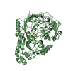
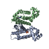
- Structure visualization
| Structure viewer | Molecule: MolmilJmol/JSmol |
|---|
- Downloads & links
-Download
| PDBx/mmCIF format | 6hlx.cif.gz | 221.2 KB | Display | PDBx/mmCIF format |
|---|---|---|---|---|
| PDB format | pdb6hlx.ent.gz | 172.3 KB | Display | PDB format |
| PDBx/mmJSON format | 6hlx.json.gz | Tree view | PDBx/mmJSON format | |
| Others |  Other downloads Other downloads |
-Validation report
| Arichive directory | https://data.pdbj.org/pub/pdb/validation_reports/hl/6hlxftp://data.pdbj.org/pub/pdb/validation_reports/hl/6hlx | HTTPS FTP |
|---|
-Related structure data
| Related structure data | 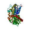 6hlyC 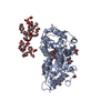 6hlzC 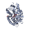 6hm2C 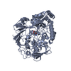 1dppS 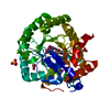 3f1qS S: Starting model for refinement C: citing same article ( |
|---|---|
| Similar structure data |


-Links
 PDBj
PDBj- Assembly
| Deposited unit | 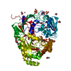
| ||||||||
|---|---|---|---|---|---|---|---|---|---|
| 1 |
| ||||||||
| Unit cell |
|
-Components
-Protein , 1 types, 1 molecules A
| #1: Protein | Mass: 54459.277 Da / Num. of mol.: 1 Source method: isolated from a genetically manipulated source Source: (gene. exp.) Rhizobium radiobacter (Agrobacterium genomosp. 4)Gene: agaA, A6U90_18825, At1D1609_52290 / Production host: |
|---|
-Non-polymers , 9 types, 507 molecules 


| #2: Chemical |  Mass: 92.094 Da / Num. of mol.: 3 / Source method: obtained synthetically / Formula: C3H8O3 Mass: 92.094 Da / Num. of mol.: 3 / Source method: obtained synthetically / Formula: C3H8O3#3: Chemical | ChemComp-G9Z / |  Mass: 291.255 Da / Num. of mol.: 1 / Source method: obtained synthetically / Formula: C11H17NO8 Mass: 291.255 Da / Num. of mol.: 1 / Source method: obtained synthetically / Formula: C11H17NO8#4: Chemical | ChemComp-MES / |  Mass: 195.237 Da / Num. of mol.: 1 / Source method: obtained synthetically / Formula: C6H13NO4S / Comment: pH buffer*YM Mass: 195.237 Da / Num. of mol.: 1 / Source method: obtained synthetically / Formula: C6H13NO4S / Comment: pH buffer*YM#5: Chemical | ChemComp-ACT /  Mass: 59.044 Da / Num. of mol.: 7 / Source method: obtained synthetically / Formula: C2H3O2 Mass: 59.044 Da / Num. of mol.: 7 / Source method: obtained synthetically / Formula: C2H3O2#6: Chemical | ChemComp-ZN /  Mass: 65.409 Da / Num. of mol.: 6 / Source method: obtained synthetically / Formula: Zn Mass: 65.409 Da / Num. of mol.: 6 / Source method: obtained synthetically / Formula: Zn#7: Chemical | ChemComp-SO4 /  Mass: 96.063 Da / Num. of mol.: 4 / Source method: obtained synthetically / Formula: SO4 Mass: 96.063 Da / Num. of mol.: 4 / Source method: obtained synthetically / Formula: SO4#8: Chemical | ChemComp-EDO /  Mass: 62.068 Da / Num. of mol.: 14 / Source method: obtained synthetically / Formula: C2H6O2 Mass: 62.068 Da / Num. of mol.: 14 / Source method: obtained synthetically / Formula: C2H6O2#9: Chemical | ChemComp-PEG / |  Mass: 106.120 Da / Num. of mol.: 1 / Source method: obtained synthetically / Formula: C4H10O3 Mass: 106.120 Da / Num. of mol.: 1 / Source method: obtained synthetically / Formula: C4H10O3#10: Water | ChemComp-HOH / | Mass: 18.015 Da / Num. of mol.: 470 / Source method: isolated from a natural source / Formula: H2O |
|---|
-Experimental details
-Experiment
| Experiment | Method: X-RAY DIFFRACTION / Number of used crystals: 1 |
|---|
- Sample preparation
| Crystal | Density Matthews: 3.02 Å3/Da / Density % sol: 59.25 % |
|---|---|
| Crystal grow | Temperature: 292 K / Method: vapor diffusion, hanging drop / Details: 1.8 M AS, 0.1 M Mes pH 6.5, 0.2 M Zn acetate |
-Data collection
| Diffraction | Mean temperature: 100 K |
|---|---|
| Diffraction source | Source: SYNCHROTRON / Site: SOLEIL  / Beamline: PROXIMA 2 / Wavelength: 0.98 Å / Beamline: PROXIMA 2 / Wavelength: 0.98 Å |
| Detector | Type: DECTRIS PILATUS 2M / Detector: PIXEL / Date: Feb 5, 2016 |
| Radiation | Protocol: SINGLE WAVELENGTH / Monochromatic (M) / Laue (L): M / Scattering type: x-ray |
| Radiation wavelength | Wavelength: 0.98 Å / Relative weight: 1 |
| Reflection | Resolution: 1.65→52.48 Å / Num. obs: 82437 / % possible obs: 99.9 % / Redundancy: 20 % / Biso Wilson estimate: 26.82 Å2 / Rsym value: 0.132 / Net I/σ(I): 19.2 |
| Reflection shell | Resolution: 1.65→1.74 Å / Rsym value: 2.052 |
- Processing
| Software |
| ||||||||||||||||||||||||||||||||||||||||||||||||||||||||||||||||||||||||||||||||||||||||||||||||||||||||||||||||||
|---|---|---|---|---|---|---|---|---|---|---|---|---|---|---|---|---|---|---|---|---|---|---|---|---|---|---|---|---|---|---|---|---|---|---|---|---|---|---|---|---|---|---|---|---|---|---|---|---|---|---|---|---|---|---|---|---|---|---|---|---|---|---|---|---|---|---|---|---|---|---|---|---|---|---|---|---|---|---|---|---|---|---|---|---|---|---|---|---|---|---|---|---|---|---|---|---|---|---|---|---|---|---|---|---|---|---|---|---|---|---|---|---|---|---|---|
| Refinement | Method to determine structure: MOLECULAR REPLACEMENT Starting model: 3F1Q and 1DPP Resolution: 1.65→52.48 Å / Cor.coef. Fo:Fc: 0.97 / Cor.coef. Fo:Fc free: 0.965 / SU R Cruickshank DPI: 0.068 / Cross valid method: THROUGHOUT / σ(F): 0 / SU R Blow DPI: 0.073 / SU Rfree Blow DPI: 0.071 / SU Rfree Cruickshank DPI: 0.067
| ||||||||||||||||||||||||||||||||||||||||||||||||||||||||||||||||||||||||||||||||||||||||||||||||||||||||||||||||||
| Displacement parameters | Biso mean: 29.65 Å2
| ||||||||||||||||||||||||||||||||||||||||||||||||||||||||||||||||||||||||||||||||||||||||||||||||||||||||||||||||||
| Refine analyze | Luzzati coordinate error obs: 0.19 Å | ||||||||||||||||||||||||||||||||||||||||||||||||||||||||||||||||||||||||||||||||||||||||||||||||||||||||||||||||||
| Refinement step | Cycle: 1 / Resolution: 1.65→52.48 Å
| ||||||||||||||||||||||||||||||||||||||||||||||||||||||||||||||||||||||||||||||||||||||||||||||||||||||||||||||||||
| Refine LS restraints |
| ||||||||||||||||||||||||||||||||||||||||||||||||||||||||||||||||||||||||||||||||||||||||||||||||||||||||||||||||||
| LS refinement shell | Resolution: 1.65→1.66 Å / Total num. of bins used: 50
| ||||||||||||||||||||||||||||||||||||||||||||||||||||||||||||||||||||||||||||||||||||||||||||||||||||||||||||||||||
| Refinement TLS params. | Method: refined / Origin x: 80.1144 Å / Origin y: -31.7036 Å / Origin z: 1.2197 Å
| ||||||||||||||||||||||||||||||||||||||||||||||||||||||||||||||||||||||||||||||||||||||||||||||||||||||||||||||||||
| Refinement TLS group | Selection details: { A|25 - A|169 A|175 - A|315 A|322 - A|512 } |
