 Movie
Movie Controller


+ Open data

- Basic information
| Entry | Database: PDB / ID: 1m27 | ||||||
|---|---|---|---|---|---|---|---|
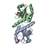
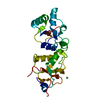
| Title | Crystal structure of SAP/FynSH3/SLAM ternary complex | ||||||
 Components Components |
| ||||||
 Keywords Keywords | SIGNALING PROTEIN / TRANSFERASE / SH2-SH3 interaction | ||||||
| Function / homology |  Function and homology information Function and homology informationnatural killer cell proliferation / negative regulation of CD40 signaling pathway / negative regulation of T cell cytokine production / regulation of vesicle fusion / myeloid dendritic cell activation involved in immune response / leukocyte chemotaxis involved in inflammatory response / negative regulation of hydrogen peroxide biosynthetic process / response to singlet oxygen / positive regulation of dendritic cell chemotaxis / positive regulation of T-helper 1 cell cytokine production ...natural killer cell proliferation / negative regulation of CD40 signaling pathway / negative regulation of T cell cytokine production / regulation of vesicle fusion / myeloid dendritic cell activation involved in immune response / leukocyte chemotaxis involved in inflammatory response / negative regulation of hydrogen peroxide biosynthetic process / response to singlet oxygen / positive regulation of dendritic cell chemotaxis / positive regulation of T-helper 1 cell cytokine production / Reelin signalling pathway / natural killer cell differentiation / perinuclear endoplasmic reticulum / NTRK2 activates RAC1 / regulation of glutamate receptor signaling pathway / heart process / negative regulation of interleukin-12 production / Activated NTRK2 signals through FYN / SEMA3A-Plexin repulsion signaling by inhibiting Integrin adhesion / regulation of calcium ion import across plasma membrane / Platelet Adhesion to exposed collagen / reelin-mediated signaling pathway / CRMPs in Sema3A signaling / FLT3 signaling through SRC family kinases / G protein-coupled glutamate receptor signaling pathway / activated T cell proliferation / positive regulation of natural killer cell mediated cytotoxicity / CD4 receptor binding / feeding behavior / positive regulation of protein localization to membrane / Nef and signal transduction / Co-stimulation by CD28 / EPH-Ephrin signaling / natural killer cell activation / cellular response to L-glutamate / DCC mediated attractive signaling / Nephrin family interactions / type 5 metabotropic glutamate receptor binding / CD28 dependent Vav1 pathway / Ephrin signaling / negative regulation of dendritic spine maintenance / positive regulation of activated T cell proliferation / dendritic spine maintenance / Regulation of KIT signaling / leukocyte migration / growth factor receptor binding / cellular response to peptide hormone stimulus / phospholipase activator activity / EPHA-mediated growth cone collapse / tau-protein kinase activity / natural killer cell mediated cytotoxicity / Co-inhibition by CTLA4 / dendrite morphogenesis / Dectin-2 family / negative regulation of T cell activation / peptide hormone receptor binding / stimulatory C-type lectin receptor signaling pathway / Fc-gamma receptor signaling pathway involved in phagocytosis / positive regulation of macrophage chemotaxis / forebrain development / PECAM1 interactions / CD8 receptor binding / response to amyloid-beta / negative regulation of type II interferon production / negative regulation of interleukin-6 production / humoral immune response / FCGR activation / Sema3A PAK dependent Axon repulsion / EPH-ephrin mediated repulsion of cells / negative regulation of tumor necrosis factor production / Role of LAT2/NTAL/LAB on calcium mobilization / cellular response to glycine / CD28 dependent PI3K/Akt signaling / ephrin receptor signaling pathway / vascular endothelial growth factor receptor signaling pathway / negative regulation of extrinsic apoptotic signaling pathway in absence of ligand / negative regulation of T cell receptor signaling pathway / regulation of immune response / positive regulation of protein targeting to membrane / cellular response to transforming growth factor beta stimulus / detection of mechanical stimulus involved in sensory perception of pain / glial cell projection / negative regulation of oxidative stress-induced intrinsic apoptotic signaling pathway / antigen binding / alpha-tubulin binding / cellular defense response / phagocytosis / postsynaptic density, intracellular component / phosphatidylinositol 3-kinase binding / ephrin receptor binding / GPVI-mediated activation cascade / T cell receptor binding / phospholipase binding / T cell costimulation / phagocytic vesicle / cellular response to platelet-derived growth factor stimulus / Signaling by ERBB2 / peptidyl-tyrosine phosphorylation / EPHB-mediated forward signaling / NCAM signaling for neurite out-growth Similarity search - Function | ||||||
| Biological species |  Homo sapiens (human) Homo sapiens (human) | ||||||
| Method |  X-RAY DIFFRACTION / MOLECULAR REPLACEMENT / Resolution: 2.5 Å X-RAY DIFFRACTION / MOLECULAR REPLACEMENT / Resolution: 2.5 Å | ||||||
 Authors Authors | Chan, B. / Griesbach, J. / Song, H.K. / Poy, F. / Terhorst, C. / Eck, M.J. | ||||||
 Citation Citation | Journal: NAT.CELL BIOL. / Year: 2003 Title: SAP couples Fyn to SLAM immune receptors. Authors: Chan, B. / Lanyi, A. / Song, H.K. / Griesbach, J. / Simarro-Grande, M. / Poy, F. / Howie, D. / Sumegi, J. / Terhorst, C. / Eck, M.J. | ||||||
| History |
|
- Structure visualization
| Structure viewer | Molecule: MolmilJmol/JSmol |
|---|
- Downloads & links
-Download
| PDBx/mmCIF format | 1m27.cif.gz | 49.4 KB | Display | PDBx/mmCIF format |
|---|---|---|---|---|
| PDB format | pdb1m27.ent.gz | 35.7 KB | Display | PDB format |
| PDBx/mmJSON format | 1m27.json.gz | Tree view | PDBx/mmJSON format | |
| Others |  Other downloads Other downloads |
-Validation report
| Arichive directory | https://data.pdbj.org/pub/pdb/validation_reports/m2/1m27ftp://data.pdbj.org/pub/pdb/validation_reports/m2/1m27 | HTTPS FTP |
|---|
-Related structure data
| Related structure data | |
|---|---|
| Similar structure data |


-Links
 PDBj
PDBj


- Assembly
| Deposited unit | 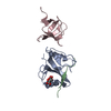
| ||||||||
|---|---|---|---|---|---|---|---|---|---|
| 1 |
| ||||||||
| Unit cell |
|
-Components
| #1: Protein | Mass: 11702.393 Da / Num. of mol.: 1 Source method: isolated from a genetically manipulated source Source: (gene. exp.) Homo sapiens (human) / Production host:  |
|---|---|
| #2: Protein/peptide | Mass: 1280.512 Da / Num. of mol.: 1 / Fragment: SLAM peptide (residues 276-286) / Source method: obtained synthetically Details: This sequence occurs naturally in humans. This sequence was chemically synthesized. References: UniProt: Q13291 |
| #3: Protein | Mass: 6938.519 Da / Num. of mol.: 1 / Fragment: SH3 domain (residues 82-143) Source method: isolated from a genetically manipulated source Source: (gene. exp.) Homo sapiens (human) / Production host: |
| #4: Chemical | ChemComp-FLC /   Mass: 189.100 Da / Num. of mol.: 1 / Source method: obtained synthetically / Formula: C6H5O7 Mass: 189.100 Da / Num. of mol.: 1 / Source method: obtained synthetically / Formula: C6H5O7 |
| #5: Water | ChemComp-HOH /  Mass: 18.015 Da / Num. of mol.: 86 / Source method: isolated from a natural source / Formula: H2O Mass: 18.015 Da / Num. of mol.: 86 / Source method: isolated from a natural source / Formula: H2O |
-Experimental details
-Experiment
| Experiment | Method: X-RAY DIFFRACTION / Number of used crystals: 1 |
|---|
- Sample preparation
| Crystal | Density Matthews: 3.21 Å3/Da / Density % sol: 61.74 % | ||||||||||||||||||||||||||||||||||||||||||||||||||||||||
|---|---|---|---|---|---|---|---|---|---|---|---|---|---|---|---|---|---|---|---|---|---|---|---|---|---|---|---|---|---|---|---|---|---|---|---|---|---|---|---|---|---|---|---|---|---|---|---|---|---|---|---|---|---|---|---|---|---|
| Crystal grow | Temperature: 297 K / Method: vapor diffusion, hanging drop / pH: 5.6 Details: Tartrate, pH 5.6, VAPOR DIFFUSION, HANGING DROP, temperature 297K | ||||||||||||||||||||||||||||||||||||||||||||||||||||||||
| Crystal grow | *PLUS Temperature: 22 ℃ / pH: 8 / Method: vapor diffusion | ||||||||||||||||||||||||||||||||||||||||||||||||||||||||
| Components of the solutions | *PLUS
|
-Data collection
| Diffraction | Mean temperature: 160 K |
|---|---|
| Diffraction source | Source: ROTATING ANODE / Type: RIGAKU RU300 / Wavelength: 1.54 Å |
| Detector | Type: MARRESEARCH / Detector: IMAGE PLATE / Date: Oct 20, 2001 / Details: Osmic Mirror |
| Radiation | Protocol: SINGLE WAVELENGTH / Monochromatic (M) / Laue (L): M / Scattering type: x-ray |
| Radiation wavelength | Wavelength: 1.54 Å / Relative weight: 1 |
| Reflection | Resolution: 2.5→20 Å / Num. all: 62846 / Num. obs: 62846 / % possible obs: 97.8 % / Observed criterion σ(I): -3 |
| Reflection shell | Resolution: 2.5→2.59 Å / % possible all: 98.4 |
| Reflection | *PLUS Lowest resolution: 20 Å / Num. obs: 9233 / % possible obs: 95.5 % / Redundancy: 3.2 % / Num. measured all: 29117 / Rmerge(I) obs: 0.044 |
| Reflection shell | *PLUS Highest resolution: 2.5 Å / Lowest resolution: 2.6 Å / % possible obs: 95.5 % / Rmerge(I) obs: 0.246 |
- Processing
| Software |
| ||||||||||||||||||||
|---|---|---|---|---|---|---|---|---|---|---|---|---|---|---|---|---|---|---|---|---|---|
| Refinement | Method to determine structure: MOLECULAR REPLACEMENT / Resolution: 2.5→20 Å / Cross valid method: THROUGHOUT / σ(F): 0 / Stereochemistry target values: Engh & Huber
| ||||||||||||||||||||
| Refine analyze |
| ||||||||||||||||||||
| Refinement step | Cycle: LAST / Resolution: 2.5→20 Å
| ||||||||||||||||||||
| Refine LS restraints |
| ||||||||||||||||||||
| LS refinement shell | Resolution: 2.5→20 Å / Rfactor Rfree error: 0.009
| ||||||||||||||||||||
| Refinement | *PLUS % reflection Rfree: 10 % / Rfactor Rfree: 0.246 / Rfactor Rwork: 0.206 | ||||||||||||||||||||
| Solvent computation | *PLUS | ||||||||||||||||||||
| Displacement parameters | *PLUS | ||||||||||||||||||||
| Refine LS restraints | *PLUS
| ||||||||||||||||||||
| LS refinement shell | *PLUS Lowest resolution: 2.6 Å |
