 Movie
Movie Controller

[English] 日本語
 Yorodumi
- PDB-1lxy: Crystal Structure of Arginine Deiminase covalently linked with L-... -
+ Open data

- Basic information
| Entry | Database: PDB / ID: 1lxy | |||||||||
|---|---|---|---|---|---|---|---|---|---|---|
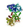
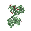
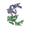
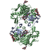
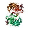
| Title | Crystal Structure of Arginine Deiminase covalently linked with L-citrulline | |||||||||
 Components Components | Arginine Deiminase | |||||||||
 Keywords Keywords | HYDROLASE / deiminase / 5-fold pseudo-symmetric domain / 5-Helix bundle domain | |||||||||
| Function / homology |  Function and homology information Function and homology informationarginine deiminase / arginine deiminase activity / L-arginine deiminase pathway / : / cytoplasm Similarity search - Function | |||||||||
| Biological species |  Mycoplasma arginini (bacteria) Mycoplasma arginini (bacteria) | |||||||||
| Method |  X-RAY DIFFRACTION / SYNCHROTRON / MIR / Resolution: 2 Å X-RAY DIFFRACTION / SYNCHROTRON / MIR / Resolution: 2 Å | |||||||||
 Authors Authors | Das, K. / Buttler, G.H. / Kwiatkowski, V. / Yadav, P. / Arnold, E. | |||||||||
 Citation Citation | Journal: Structure / Year: 2004 Title: Crystal structures of arginine deiminase with covalent reaction intermediates; implications for catalytic mechanism Authors: Das, K. / Buttler, G.H. / Kwiatkowski, V. / Clark Jr., A.D. / Yadav, P. / Arnold, E. | |||||||||
| History |
|
- Structure visualization
| Structure viewer | Molecule: MolmilJmol/JSmol |
|---|
- Downloads & links
-Download
| PDBx/mmCIF format | 1lxy.cif.gz | 183.6 KB | Display | PDBx/mmCIF format |
|---|---|---|---|---|
| PDB format | pdb1lxy.ent.gz | 146.2 KB | Display | PDB format |
| PDBx/mmJSON format | 1lxy.json.gz | Tree view | PDBx/mmJSON format | |
| Others |  Other downloads Other downloads |
-Validation report
| Summary document | 1lxy_validation.pdf.gz | 461.7 KB | Display | wwPDB validaton report |
|---|---|---|---|---|
| Full document | 1lxy_full_validation.pdf.gz | 469.6 KB | Display | |
| Data in XML | 1lxy_validation.xml.gz | 36.3 KB | Display | |
| Data in CIF | 1lxy_validation.cif.gz | 53.4 KB | Display | |
| Arichive directory | https://data.pdbj.org/pub/pdb/validation_reports/lx/1lxyftp://data.pdbj.org/pub/pdb/validation_reports/lx/1lxy | HTTPS FTP |
-Related structure data


-Links
 PDBj
PDBj- Assembly
| Deposited unit | 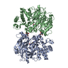
| ||||||||
|---|---|---|---|---|---|---|---|---|---|
| 1 |
| ||||||||
| Unit cell |
|
-Components
| #1: Protein | Mass: 46431.973 Da / Num. of mol.: 2 / Source method: isolated from a natural source Details: 5-fold pseudosymmetric guanidino group modifying enzyme Source: (natural) Mycoplasma arginini (bacteria) / References: UniProt: P23793, arginine deiminase#2: Chemical |   Type: L-peptide linking / Mass: 175.186 Da / Num. of mol.: 2 / Source method: obtained synthetically / Formula: C6H13N3O3 Type: L-peptide linking / Mass: 175.186 Da / Num. of mol.: 2 / Source method: obtained synthetically / Formula: C6H13N3O3#3: Chemical |   Mass: 122.143 Da / Num. of mol.: 2 / Source method: obtained synthetically / Formula: C4H12NO3 / Comment: pH buffer*YM Mass: 122.143 Da / Num. of mol.: 2 / Source method: obtained synthetically / Formula: C4H12NO3 / Comment: pH buffer*YM#4: Water | ChemComp-HOH / |  Mass: 18.015 Da / Num. of mol.: 550 / Source method: isolated from a natural source / Formula: H2O Mass: 18.015 Da / Num. of mol.: 550 / Source method: isolated from a natural source / Formula: H2OHas protein modification | Y | |
|---|
-Experimental details
-Experiment
| Experiment | Method: X-RAY DIFFRACTION / Number of used crystals: 1 |
|---|
- Sample preparation
| Crystal | Density Matthews: 2.31 Å3/Da / Density % sol: 46.2 % | ||||||||||||||||||||||||||||||
|---|---|---|---|---|---|---|---|---|---|---|---|---|---|---|---|---|---|---|---|---|---|---|---|---|---|---|---|---|---|---|---|
| Crystal grow | Temperature: 282 K / Method: vapor diffusion, hanging drop / pH: 6.5 Details: PEG 6000, Potassium phosphate, L-citrulline, pH 6.5, VAPOR DIFFUSION, HANGING DROP at 282K | ||||||||||||||||||||||||||||||
| Crystal grow | *PLUS Temperature: 4 ℃ / pH: 7 / Method: vapor diffusion, hanging drop | ||||||||||||||||||||||||||||||
| Components of the solutions | *PLUS
|
-Data collection
| Diffraction |
| ||||||||||||||||||
|---|---|---|---|---|---|---|---|---|---|---|---|---|---|---|---|---|---|---|---|
| Diffraction source |
| ||||||||||||||||||
| Detector |
| ||||||||||||||||||
| Radiation | Protocol: SINGLE WAVELENGTH / Monochromatic (M) / Laue (L): M / Scattering type: x-ray | ||||||||||||||||||
| Radiation wavelength |
| ||||||||||||||||||
| Reflection | Resolution: 2→99 Å / Num. all: 59662 / Num. obs: 59662 / % possible obs: 97.1 % / Observed criterion σ(I): 0 / Rmerge(I) obs: 0.083 / Net I/σ(I): 11.2 | ||||||||||||||||||
| Reflection shell | Resolution: 2→2.03 Å / Rmerge(I) obs: 0.219 / Mean I/σ(I) obs: 4.6 / % possible all: 90.4 | ||||||||||||||||||
| Reflection | *PLUS Highest resolution: 2 Å / Redundancy: 3.37 % | ||||||||||||||||||
| Reflection shell | *PLUS % possible obs: 90.4 % / Rmerge(I) obs: 0.22 |

- Processing
| Software |
| ||||||||||||||||||||||||||||||||||||
|---|---|---|---|---|---|---|---|---|---|---|---|---|---|---|---|---|---|---|---|---|---|---|---|---|---|---|---|---|---|---|---|---|---|---|---|---|---|
| Refinement | Method to determine structure: MIR / Resolution: 2→19.95 Å / Rfactor Rfree error: 0.004 / Data cutoff high absF: 1799833.91 / Data cutoff low absF: 0 / Isotropic thermal model: RESTRAINED / Cross valid method: THROUGHOUT / σ(F): 1 / Stereochemistry target values: Engh & Huber
| ||||||||||||||||||||||||||||||||||||
| Solvent computation | Solvent model: FLAT MODEL / Bsol: 47.548 Å2 / ksol: 0.361371 e/Å3 | ||||||||||||||||||||||||||||||||||||
| Displacement parameters | Biso mean: 23.9 Å2
| ||||||||||||||||||||||||||||||||||||
| Refine analyze |
| ||||||||||||||||||||||||||||||||||||
| Refinement step | Cycle: LAST / Resolution: 2→19.95 Å
| ||||||||||||||||||||||||||||||||||||
| Refine LS restraints |
| ||||||||||||||||||||||||||||||||||||
| LS refinement shell | Resolution: 2→2.13 Å / Rfactor Rfree error: 0.015 / Total num. of bins used: 6
| ||||||||||||||||||||||||||||||||||||
| Xplor file |
| ||||||||||||||||||||||||||||||||||||
| Refinement | *PLUS Highest resolution: 2 Å / Lowest resolution: 20 Å / Rfactor all: 4.8 | ||||||||||||||||||||||||||||||||||||
| Solvent computation | *PLUS | ||||||||||||||||||||||||||||||||||||
| Displacement parameters | *PLUS | ||||||||||||||||||||||||||||||||||||
| Refine LS restraints | *PLUS
|
