dimethylargininase / dimethylargininase activity / : / arginine metabolic process / amino acid binding / positive regulation of nitric oxide biosynthetic process / metal ion binding Similarity search - Function
Mass: 18.015 Da / Num. of mol.: 144 / Source method: isolated from a natural source / Formula: H2O
Compound details
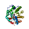
CHAIN A ENGINEERED MUTATION CYS249SER
Sequence details
PHE A 0 CLONING ARTIFACT
-
Experimental details
-
Experiment
Experiment
Method: X-RAY DIFFRACTION / Number of used crystals: 1
-
Sample preparation
Crystal
Density Matthews: 2.1 Å3/Da / Density % sol: 40 %
Crystal grow
Method: vapor diffusion, sitting drop / pH: 8.5 Details: SITTING DROPS; EQUAL QUANTITIES OF PROTEIN SOLUTION AND WELL SOLUTION. PROTEIN SOLUTION 14 MG/ML IN 50MM TRIS PH8, 5MMDTT, 10:1 MOLAR RATIO OF CITRULLINE. WEEL SOLUTION ).1M TRIS PH 8.5, 0. ...Details: SITTING DROPS; EQUAL QUANTITIES OF PROTEIN SOLUTION AND WELL SOLUTION. PROTEIN SOLUTION 14 MG/ML IN 50MM TRIS PH8, 5MMDTT, 10:1 MOLAR RATIO OF CITRULLINE. WEEL SOLUTION ).1M TRIS PH 8.5, 0.2M NA ACETATE, 25%W/V PEG4000.
 Movie
Movie Controller

 Yorodumi
 Open data
 Basic information
 Components
 Keywords
 Function and homology information
 PSEUDOMONAS AERUGINOSA (bacteria)
 X-RAY DIFFRACTION /
  Authors
 Citation
 Structure visualization
 Downloads & links
 Other downloads
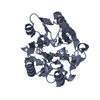


 PDBj
PDBj Assembly

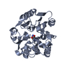
 Type: L-peptide linking / Mass: 175.186 Da / Num. of mol.: 1 / Source method: obtained synthetically / Formula: C6H13N3O3
 Mass: 18.015 Da / Num. of mol.: 144 / Source method: isolated from a natural source / Formula: H2O
 Sample preparation
 / Beamline: PX14.1 / Wavelength: 1.244
  Processing