 Movie
Movie Controller

[English] 日本語
 Yorodumi
- PDB-1lt1: SLIDING HELIX INDUCED CHANGE OF COORDINATION GEOMETRY IN A MODEL ... -
+ Open data

- Basic information
| Entry | Database: PDB / ID: 1lt1 | ||||||
|---|---|---|---|---|---|---|---|
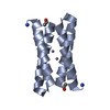
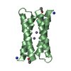
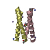
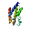
| Title | SLIDING HELIX INDUCED CHANGE OF COORDINATION GEOMETRY IN A MODEL DI-MN(II) PROTEIN | ||||||
 Components Components | L13G-DF1 | ||||||
 Keywords Keywords | DE NOVO PROTEIN / ALPHA-HELICAL BUNDLE / PROTEIN DESIGN / SLIDING HELIX | ||||||
| Function / homology | Immunoglobulin FC, subunit C / Single alpha-helices involved in coiled-coils or other helix-helix interfaces / Up-down Bundle / Mainly Alpha / :  Function and homology information Function and homology information | ||||||
| Method |  X-RAY DIFFRACTION / SYNCHROTRON / MOLECULAR REPLACEMENT / Resolution: 1.91 Å X-RAY DIFFRACTION / SYNCHROTRON / MOLECULAR REPLACEMENT / Resolution: 1.91 Å | ||||||
 Authors Authors | Di Costanzo, L. / Geremia, S. | ||||||
 Citation Citation | Journal: Angew.Chem.Int.Ed.Engl. / Year: 2003 Title: Sliding helix and change of coordination geometry in a model di-MnII protein Authors: Degrado, W.F. / Di Costanzo, L. / Geremia, S. / Lombardi, A. / Pavone, V. / Randaccio, L. #1: Journal: J.Am.Chem.Soc. / Year: 2001Title: Toward the De Novo Design of a Catalytically Active Helix-Bundle: A Substrate-accessible Carboxylate-Bridged Dinuclear Metal Center Authors: Di Costanzo, L. / Wade, H. / Geremia, S. / Randaccio, L. / Pavone, V. / Degrado, W.F. / Lombardi, A. #2: Journal: Proc.Natl.Acad.Sci.USA / Year: 2000Title: Inaugural article: Retrostructural Analysis of Metalloproteins: Application to the Design of a Minimal Model for Diiron Proteins Authors: Lombardi, A. / Summa, C.M. / Geremia, S. / Randaccio, L. / Pavone, V. / Degrado, W.F. | ||||||
| History |
|
- Structure visualization
| Structure viewer | Molecule: MolmilJmol/JSmol |
|---|
- Downloads & links
-Download
| PDBx/mmCIF format | 1lt1.cif.gz | 100.3 KB | Display | PDBx/mmCIF format |
|---|---|---|---|---|
| PDB format | pdb1lt1.ent.gz | 78.3 KB | Display | PDB format |
| PDBx/mmJSON format | 1lt1.json.gz | Tree view | PDBx/mmJSON format | |
| Others |  Other downloads Other downloads |
-Validation report
| Arichive directory | https://data.pdbj.org/pub/pdb/validation_reports/lt/1lt1ftp://data.pdbj.org/pub/pdb/validation_reports/lt/1lt1 | HTTPS FTP |
|---|
-Related structure data
| Related structure data |  1jm0S S: Starting model for refinement |
|---|---|
| Similar structure data |


-Links
 PDBj
PDBj
- Assembly
| Deposited unit | 
| ||||||||
|---|---|---|---|---|---|---|---|---|---|
| 1 | 
| ||||||||
| 2 | 
| ||||||||
| 3 | 
| ||||||||
| 4 | 
| ||||||||
| Unit cell |
|
-Components
| #1: Protein/peptide | Mass: 5814.788 Da / Num. of mol.: 8 / Source method: obtained synthetically / Details: This peptide was chemically synthetized. #2: Chemical | ChemComp-MN /   Mass: 54.938 Da / Num. of mol.: 12 / Source method: obtained synthetically / Formula: Mn Mass: 54.938 Da / Num. of mol.: 12 / Source method: obtained synthetically / Formula: Mn#3: Water | ChemComp-HOH / |  Mass: 18.015 Da / Num. of mol.: 290 / Source method: isolated from a natural source / Formula: H2O Mass: 18.015 Da / Num. of mol.: 290 / Source method: isolated from a natural source / Formula: H2OHas protein modification | Y | |
|---|
-Experimental details
-Experiment
| Experiment | Method: X-RAY DIFFRACTION / Number of used crystals: 1 |
|---|
- Sample preparation
| Crystal | Density Matthews: 2.38 Å3/Da / Density % sol: 42 % | |||||||||||||||||||||||||||||||||||
|---|---|---|---|---|---|---|---|---|---|---|---|---|---|---|---|---|---|---|---|---|---|---|---|---|---|---|---|---|---|---|---|---|---|---|---|---|
| Crystal grow | Temperature: 277 K / Method: vapor diffusion, hanging drop / pH: 7.5 Details: PEG400, MANGANESE ACETATE, BUFFER TRIS, pH 7.5, VAPOR DIFFUSION, HANGING DROP, temperature 277K | |||||||||||||||||||||||||||||||||||
| Crystal grow | *PLUS | |||||||||||||||||||||||||||||||||||
| Components of the solutions | *PLUS
|
-Data collection
| Diffraction | Mean temperature: 100 K |
|---|---|
| Diffraction source | Source: SYNCHROTRON / Site: ELETTRA  / Beamline: 5.2R / Wavelength: 1.2 / Wavelength: 1.2 Å / Beamline: 5.2R / Wavelength: 1.2 / Wavelength: 1.2 Å |
| Detector | Type: MARRESEARCH / Detector: IMAGE PLATE / Details: MIRROR |
| Radiation | Monochromator: SI(111) / Protocol: SINGLE WAVELENGTH / Monochromatic (M) / Laue (L): M / Scattering type: x-ray |
| Radiation wavelength | Wavelength: 1.2 Å / Relative weight: 1 |
| Reflection | Resolution: 1.91→6.03 Å / Num. all: 38634 / Num. obs: 38634 / % possible obs: 96.6 % / Redundancy: 2.5 % / Biso Wilson estimate: 16.7 Å2 / Rmerge(I) obs: 0.125 / Rsym value: 0.125 / Net I/σ(I): 6.9 |
| Reflection shell | Resolution: 1.91→2.01 Å / Redundancy: 2.4 % / Rmerge(I) obs: 0.463 / Mean I/σ(I) obs: 2.1 / Num. unique all: 5184 / Rsym value: 0.463 / % possible all: 96.6 |
| Reflection | *PLUS Num. measured all: 94932 |
- Processing
| Software |
| ||||||||||||||||||||
|---|---|---|---|---|---|---|---|---|---|---|---|---|---|---|---|---|---|---|---|---|---|
| Refinement | Method to determine structure: MOLECULAR REPLACEMENT Starting model: AB DIMER FROM THE STRUCTURE 1JM0 Resolution: 1.91→20 Å / Cross valid method: THROUGHOUT / σ(F): 0
| ||||||||||||||||||||
| Displacement parameters | Biso mean: 21.179 Å2
| ||||||||||||||||||||
| Refinement step | Cycle: LAST / Resolution: 1.91→20 Å
| ||||||||||||||||||||
| Refine LS restraints |
| ||||||||||||||||||||
| Refinement | *PLUS % reflection Rfree: 5 % / Rfactor Rfree: 0.244 / Rfactor Rwork: 0.201 | ||||||||||||||||||||
| Solvent computation | *PLUS | ||||||||||||||||||||
| Displacement parameters | *PLUS | ||||||||||||||||||||
| Refine LS restraints | *PLUS
|
