 Movie
Movie Controller


+ Open data

- Basic information
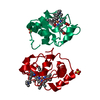
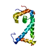
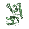
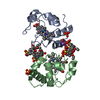
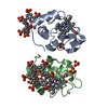
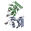
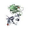
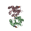
| Entry | Database: PDB / ID: 1ln0 | ||||||
|---|---|---|---|---|---|---|---|
| Title | Structure of the Catalytic Domain of Homing Endonuclease I-TevI | ||||||
 Components Components | intron-associated endonuclease 1 | ||||||
 Keywords Keywords | HYDROLASE / alpha/beta fold | ||||||
| Function / homology |  Function and homology information Function and homology informationdouble-stranded DNA endonuclease activity / intron homing / DNA-binding transcription repressor activity / transcription repressor complex / endonuclease activity / sequence-specific DNA binding / Hydrolases; Acting on ester bonds / negative regulation of DNA-templated transcription / DNA binding / zinc ion binding Similarity search - Function | ||||||
| Biological species |  Enterobacteria phage T4 (virus) Enterobacteria phage T4 (virus) | ||||||
| Method |  X-RAY DIFFRACTION / SYNCHROTRON / SIRAS / Resolution: 2 Å X-RAY DIFFRACTION / SYNCHROTRON / SIRAS / Resolution: 2 Å | ||||||
 Authors Authors | Van Roey, P. / Meehan, L. / Kowalski, J.C. / Belfort, M. / Derbyshire, V. | ||||||
 Citation Citation | Journal: Nat.Struct.Biol. / Year: 2002 Title: Catalytic domain structure and hypothesis for function of GIY-YIG intron endonuclease I-TevI. Authors: Van Roey, P. / Meehan, L. / Kowalski, J.C. / Belfort, M. / Derbyshire, V. | ||||||
| History |
|
- Structure visualization
| Structure viewer | Molecule: MolmilJmol/JSmol |
|---|
- Downloads & links
-Download
| PDBx/mmCIF format | 1ln0.cif.gz | 54.8 KB | Display | PDBx/mmCIF format |
|---|---|---|---|---|
| PDB format | pdb1ln0.ent.gz | 40 KB | Display | PDB format |
| PDBx/mmJSON format | 1ln0.json.gz | Tree view | PDBx/mmJSON format | |
| Others |  Other downloads Other downloads |
-Validation report
| Arichive directory | https://data.pdbj.org/pub/pdb/validation_reports/ln/1ln0ftp://data.pdbj.org/pub/pdb/validation_reports/ln/1ln0 | HTTPS FTP |
|---|
-Related structure data

-Links
 PDBj
PDBj- Assembly
| Deposited unit | 
| ||||||||
|---|---|---|---|---|---|---|---|---|---|
| 1 |
| ||||||||
| Unit cell |
| ||||||||
| Details | dimer of domain covalently linked by disulfide bridge between Cys39 residues |
-Components
| #1: Protein | Mass: 11377.977 Da / Num. of mol.: 2 / Fragment: catalytic domain (residues 1 to 97) / Mutation: R27A Source method: isolated from a genetically manipulated source Source: (gene. exp.) Enterobacteria phage T4 (virus) / Genus: T4-like viruses / Species: Enterobacteria phage T4 sensu lato / Production host:  References: UniProt: P13299, Hydrolases; Acting on ester bonds #2: Chemical |   Mass: 96.063 Da / Num. of mol.: 2 / Source method: obtained synthetically / Formula: SO4 Mass: 96.063 Da / Num. of mol.: 2 / Source method: obtained synthetically / Formula: SO4#3: Water | ChemComp-HOH / |  Mass: 18.015 Da / Num. of mol.: 208 / Source method: isolated from a natural source / Formula: H2O Mass: 18.015 Da / Num. of mol.: 208 / Source method: isolated from a natural source / Formula: H2OHas protein modification | Y | |
|---|
-Experimental details
-Experiment
| Experiment | Method: X-RAY DIFFRACTION / Number of used crystals: 2 |
|---|
- Sample preparation
| Crystal | Density Matthews: 2.73 Å3/Da / Density % sol: 54.93 % | |||||||||||||||||||||||||||||||||||||||||||||||||||||||||||||||
|---|---|---|---|---|---|---|---|---|---|---|---|---|---|---|---|---|---|---|---|---|---|---|---|---|---|---|---|---|---|---|---|---|---|---|---|---|---|---|---|---|---|---|---|---|---|---|---|---|---|---|---|---|---|---|---|---|---|---|---|---|---|---|---|---|
| Crystal grow | Temperature: 283 K / Method: vapor diffusion, hanging drop / pH: 6.5 Details: 22% PEG8000, 0.1 M MES, 5% ammonium sulfate, pH 6.5, VAPOR DIFFUSION, HANGING DROP, temperature 283K | |||||||||||||||||||||||||||||||||||||||||||||||||||||||||||||||
| Crystal grow | *PLUS Temperature: 4 ℃ / pH: 7.5 | |||||||||||||||||||||||||||||||||||||||||||||||||||||||||||||||
| Components of the solutions | *PLUS
|
-Data collection
| Diffraction | Mean temperature: 100 K |
|---|---|
| Diffraction source | Source: SYNCHROTRON / Site: NSLS  / Beamline: X12C / Wavelength: 1 Å / Beamline: X12C / Wavelength: 1 Å |
| Detector | Type: BRANDEIS - B1.2 / Detector: CCD / Date: Aug 1, 2001 |
| Radiation | Protocol: SINGLE WAVELENGTH / Monochromatic (M) / Laue (L): M / Scattering type: x-ray |
| Radiation wavelength | Wavelength: 1 Å / Relative weight: 1 |
| Reflection | Resolution: 2→50 Å / Num. all: 16242 / Num. obs: 15982 / % possible obs: 92.2 % / Observed criterion σ(F): 0 / Observed criterion σ(I): 0 / Redundancy: 3.73 % / Rmerge(I) obs: 0.034 / Net I/σ(I): 33.4 |
| Reflection shell | Resolution: 2→2.1 Å / Rmerge(I) obs: 0.124 / Mean I/σ(I) obs: 8.3 / % possible all: 61.6 |
| Reflection | *PLUS Lowest resolution: 50 Å / Num. obs: 16242 |
| Reflection shell | *PLUS % possible obs: 61.6 % |
- Processing
| Software |
| |||||||||||||||||||||||||
|---|---|---|---|---|---|---|---|---|---|---|---|---|---|---|---|---|---|---|---|---|---|---|---|---|---|---|
| Refinement | Method to determine structure: SIRAS / Resolution: 2→50 Å / σ(F): 0 / σ(I): 0 / Stereochemistry target values: Engh & Huber
| |||||||||||||||||||||||||
| Refinement step | Cycle: LAST / Resolution: 2→50 Å
| |||||||||||||||||||||||||
| Refine LS restraints |
| |||||||||||||||||||||||||
| Refinement | *PLUS | |||||||||||||||||||||||||
| Solvent computation | *PLUS | |||||||||||||||||||||||||
| Displacement parameters | *PLUS | |||||||||||||||||||||||||
| Refine LS restraints | *PLUS Type: c_angle_deg / Dev ideal: 1.2 |
