| Entry | Database: PDB / ID: 5mtn
|
|---|
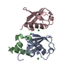
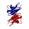
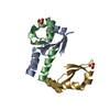
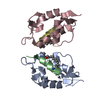
| Title | Monobody Mb(Lck_1) bound to Lck-Sh2 |
|---|
 Components Components | - Monobody Mb(Lck_1)
- Tyrosine-protein kinase Lck
|
|---|
 Keywords Keywords | TRANSFERASE / Src homology 2 / Monobodies / Directed evolution |
|---|
| Function / homology |  Function and homology information Function and homology information
regulation of lymphocyte activation / positive regulation of leukocyte cell-cell adhesion / CD27 signaling pathway / regulation of regulatory T cell differentiation / gamma-delta T cell differentiation / positive regulation of gamma-delta T cell differentiation / Fc-gamma receptor signaling pathway / FLT3 signaling through SRC family kinases / protein antigen binding / positive regulation of T cell receptor signaling pathway ...regulation of lymphocyte activation / positive regulation of leukocyte cell-cell adhesion / CD27 signaling pathway / regulation of regulatory T cell differentiation / gamma-delta T cell differentiation / positive regulation of gamma-delta T cell differentiation / Fc-gamma receptor signaling pathway / FLT3 signaling through SRC family kinases / protein antigen binding / positive regulation of T cell receptor signaling pathway / Nef Mediated CD4 Down-regulation / CD4 receptor binding / Nef and signal transduction / Co-stimulation by CD28 / Interleukin-2 signaling / positive regulation of heterotypic cell-cell adhesion / CD28 dependent Vav1 pathway / leukocyte migration / Regulation of KIT signaling / phospholipase activator activity / Co-inhibition by CTLA4 / Translocation of ZAP-70 to Immunological synapse / Phosphorylation of CD3 and TCR zeta chains / PECAM1 interactions / CD8 receptor binding / pericentriolar material / protein serine/threonine phosphatase activity / Generation of second messenger molecules / hemopoiesis / T cell differentiation / Co-inhibition by PD-1 / RHOH GTPase cycle / immunological synapse / CD28 dependent PI3K/Akt signaling / phosphatidylinositol 3-kinase binding / GPVI-mediated activation cascade / T cell receptor binding / phospholipase binding / T cell costimulation / phosphotyrosine residue binding / positive regulation of intrinsic apoptotic signaling pathway / release of sequestered calcium ion into cytosol / SH2 domain binding / cell surface receptor protein tyrosine kinase signaling pathway / Signaling by phosphorylated juxtamembrane, extracellular and kinase domain KIT mutants / T cell activation / B cell receptor signaling pathway / non-specific protein-tyrosine kinase / non-membrane spanning protein tyrosine kinase activity / Signaling by SCF-KIT / platelet activation / positive regulation of T cell activation / Constitutive Signaling by Aberrant PI3K in Cancer / cell-cell junction / DAP12 signaling / PIP3 activates AKT signaling / Downstream TCR signaling / T cell receptor signaling pathway / ATPase binding / PI5P, PP2A and IER3 Regulate PI3K/AKT Signaling / protein tyrosine kinase activity / protein phosphatase binding / intracellular signal transduction / membrane raft / response to xenobiotic stimulus / signaling receptor binding / positive regulation of gene expression / protein kinase binding / extracellular exosome / ATP binding / identical protein binding / plasma membrane / cytoplasm / cytosolSimilarity search - Function Lck, SH3 domain / Tyrosine-protein kinase Lck, SH2 domain / SH2 domain / SHC Adaptor Protein / : / SH3 domain / SH2 domain / Src homology 2 (SH2) domain profile. / Src homology 2 domains / SH2 domain ...Lck, SH3 domain / Tyrosine-protein kinase Lck, SH2 domain / SH2 domain / SHC Adaptor Protein / : / SH3 domain / SH2 domain / Src homology 2 (SH2) domain profile. / Src homology 2 domains / SH2 domain / Src homology 3 domains / SH2 domain superfamily / SH3-like domain superfamily / Src homology 3 (SH3) domain profile. / SH3 domain / Tyrosine-protein kinase, catalytic domain / Tyrosine kinase, catalytic domain / Tyrosine protein kinases specific active-site signature. / Tyrosine-protein kinase, active site / Protein tyrosine and serine/threonine kinase / Serine-threonine/tyrosine-protein kinase, catalytic domain / Protein kinase, ATP binding site / Protein kinases ATP-binding region signature. / Immunoglobulins / Protein kinase domain profile. / Protein kinase domain / Protein kinase-like domain superfamily / Immunoglobulin-like / Sandwich / 2-Layer Sandwich / Mainly Beta / Alpha BetaSimilarity search - Domain/homology |
|---|
| Biological species |  Homo sapiens (human) Homo sapiens (human)
Synthetic construct (others) |
|---|
| Method |  X-RAY DIFFRACTION / SYNCHROTRON / MOLECULAR REPLACEMENT / Resolution: 2.85 Å X-RAY DIFFRACTION / SYNCHROTRON / MOLECULAR REPLACEMENT / Resolution: 2.85 Å |
|---|
 Authors Authors | Pojer, F. / Kukenshoner, T. / Koide, S. / Hantschel, O. |
|---|
 Citation Citation | Journal: J. Mol. Biol. / Year: 2017
Title: Selective Targeting of SH2 Domain-Phosphotyrosine Interactions of Src Family Tyrosine Kinases with Monobodies.
Authors: Kukenshoner, T. / Schmit, N.E. / Bouda, E. / Sha, F. / Pojer, F. / Koide, A. / Seeliger, M. / Koide, S. / Hantschel, O. |
|---|
| History | | Deposition | Jan 10, 2017 | Deposition site: PDBE / Processing site: PDBE |
|---|
| Revision 1.0 | Apr 5, 2017 | Provider: repository / Type: Initial release |
|---|
| Revision 1.1 | May 3, 2017 | Group: Database references |
|---|
| Revision 1.2 | Oct 16, 2019 | Group: Data collection / Category: reflns_shell |
|---|
| Revision 1.3 | May 8, 2024 | Group: Data collection / Database references / Category: chem_comp_atom / chem_comp_bond / database_2
Item: _database_2.pdbx_DOI / _database_2.pdbx_database_accession |
|---|
|
|---|
 Movie
Movie Controller


 Open data
 Basic information
 Structure visualization
 Downloads & links
 Other downloads
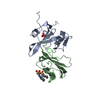


 PDBj
PDBj


 Assembly
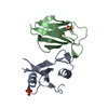


 Mass: 96.063 Da / Num. of mol.: 3 / Source method: obtained synthetically / Formula: SO4
 Sample preparation
 / Beamline: X06DA / Wavelength: 1 Å
 Processing