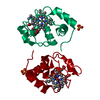
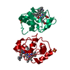
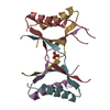
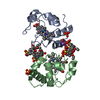
Release of apoptotic factors from the mitochondria / Pyroptosis / Detoxification of Reactive Oxygen Species / Respiratory electron transport / cardiolipin binding / mitochondrial electron transport, cytochrome c to oxygen / mitochondrial electron transport, ubiquinol to cytochrome c / mitochondrial intermembrane space / electron transfer activity / heme binding ...Release of apoptotic factors from the mitochondria / Pyroptosis / Detoxification of Reactive Oxygen Species / Respiratory electron transport / cardiolipin binding / mitochondrial electron transport, cytochrome c to oxygen / mitochondrial electron transport, ubiquinol to cytochrome c / mitochondrial intermembrane space / electron transfer activity / heme binding / mitochondrion / metal ion binding Similarity search - Function
Cytochrome c, class IA/ IB / Cytochrome c / Cytochrome c family profile. / Cytochrome c-like domain / Cytochrome c-like domain superfamily Similarity search - Domain/homology
Protocol: SINGLE WAVELENGTH / Monochromatic (M) / Laue (L): M / Scattering type: x-ray
Radiation wavelength
Wavelength: 0.9786 Å / Relative weight: 1
Reflection
Resolution: 1.8→69.34 Å / Num. obs: 30379 / % possible obs: 100 % / Redundancy: 9.1 % / Biso Wilson estimate: 20 Å2 / CC1/2: 0.998 / Rmerge(I) obs: 0.12 / Net I/σ(I): 13.1
Reflection shell
Resolution: 1.8→1.84 Å / Redundancy: 8.7 % / Rmerge(I) obs: 1.9 / CC1/2: 0.459 / % possible all: 99.9
-
Phasing
Phasing
Method: molecular replacement
-
Processing
Software
Name
Version
Classification
MOSFLM
datacollection
Aimless
datascaling
REFMAC
5.8.0155
refinement
PDB_EXTRACT
3.2
dataextraction
PHASER
phasing
Refinement
Method to determine structure: MOLECULAR REPLACEMENT / Resolution: 1.8→52.78 Å / Cor.coef. Fo:Fc: 0.967 / Cor.coef. Fo:Fc free: 0.954 / SU B: 4.852 / SU ML: 0.072 / SU R Cruickshank DPI: 0.1022 / Cross valid method: THROUGHOUT / σ(F): 0 / ESU R: 0.102 / ESU R Free: 0.101 Details: HYDROGENS HAVE BEEN ADDED IN THE RIDING POSITIONS U VALUES : WITH TLS ADDED
Rfactor
Num. reflection
% reflection
Selection details
Rfree
0.1994
1595
5.3 %
RANDOM
Rwork
0.1685
-
-
-
obs
0.1701
28686
99.97 %
-
Solvent computation
Ion probe radii: 0.8 Å / Shrinkage radii: 0.8 Å / VDW probe radii: 1.2 Å
In the structure databanks used in Yorodumi, some data are registered as the other names, "COVID-19 virus" and "2019-nCoV". Here are the details of the virus and the list of structure data.
Jan 31, 2019. EMDB accession codes are about to change! (news from PDBe EMDB page)
EMDB accession codes are about to change! (news from PDBe EMDB page)
The allocation of 4 digits for EMDB accession codes will soon come to an end. Whilst these codes will remain in use, new EMDB accession codes will include an additional digit and will expand incrementally as the available range of codes is exhausted. The current 4-digit format prefixed with “EMD-” (i.e. EMD-XXXX) will advance to a 5-digit format (i.e. EMD-XXXXX), and so on. It is currently estimated that the 4-digit codes will be depleted around Spring 2019, at which point the 5-digit format will come into force.
The EM Navigator/Yorodumi systems omit the EMD- prefix.
Related info.:Q: What is EMD? / ID/Accession-code notation in Yorodumi/EM Navigator
Yorodumi is a browser for structure data from EMDB, PDB, SASBDB, etc.
This page is also the successor to EM Navigator detail page, and also detail information page/front-end page for Omokage search.
The word "yorodu" (or yorozu) is an old Japanese word meaning "ten thousand". "mi" (miru) is to see.
Related info.:EMDB / PDB / SASBDB / Comparison of 3 databanks / Yorodumi Search / Aug 31, 2016. New EM Navigator & Yorodumi / Yorodumi Papers / Jmol/JSmol / Function and homology information / Changes in new EM Navigator and Yorodumi
 Movie
Movie Controller


 Open data
 Basic information
 Components
 Keywords
 Function and homology information
 X-RAY DIFFRACTION /
  Authors
 Citation
 Structure visualization
 Downloads & links
 Other downloads
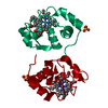


 PDBj
PDBj


 Assembly
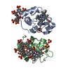


 Mass: 618.503 Da / Num. of mol.: 2 / Source method: obtained synthetically / Formula: C34H34FeN4O4
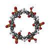
 Mass: 1116.611 Da / Num. of mol.: 2 / Source method: obtained synthetically / Formula: C42H42O24P6
 Mass: 96.063 Da / Num. of mol.: 5 / Source method: obtained synthetically / Formula: SO4
 Mass: 18.015 Da / Num. of mol.: 180 / Source method: isolated from a natural source / Formula: H2O
 Sample preparation
 / Beamline: PROXIMA 2 / Wavelength: 0.9786 Å
 Processing