 Movie
Movie Controller


+ Open data

- Basic information
| Entry | Database: PDB / ID: 1lcs | |||||||||
|---|---|---|---|---|---|---|---|---|---|---|
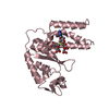
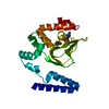
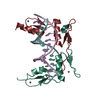
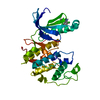
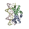
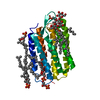
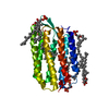
| Title | RECEPTOR-BINDING DOMAIN FROM SUBGROUP B FELINE LEUKEMIA VIRUS | |||||||||
 Components Components | FELINE LEUKEMIA VIRUS RECEPTOR-BINDING DOMAIN | |||||||||
 Keywords Keywords | VIRAL PROTEIN / antiparallel beta-sandwich glycoprotein | |||||||||
| Function / homology |  Function and homology information Function and homology informationfusion of virus membrane with host plasma membrane / viral envelope / symbiont entry into host cell / virion attachment to host cell / host cell plasma membrane / virion membrane Similarity search - Function | |||||||||
| Biological species |  Feline leukemia virus Feline leukemia virus | |||||||||
| Method |  X-RAY DIFFRACTION / SYNCHROTRON / MOLECULAR REPLACEMENT / Resolution: 2.5 Å X-RAY DIFFRACTION / SYNCHROTRON / MOLECULAR REPLACEMENT / Resolution: 2.5 Å | |||||||||
 Authors Authors | Barnett, A.L. / Wensel, D.L. / Li, W. / Fass, D. / Cunningham, J.M. | |||||||||
 Citation Citation | Journal: J.Virol. / Year: 2003 Title: Structure and Mechanism of a Coreceptor for Infection by a pathogenic feline retrovirus Authors: Barnett, A.L. / Wensel, D.L. / Li, W. / Fass, D. / Cunningham, J.M. | |||||||||
| History |
|
- Structure visualization
| Structure viewer | Molecule: MolmilJmol/JSmol |
|---|
- Downloads & links
-Download
| PDBx/mmCIF format | 1lcs.cif.gz | 102.5 KB | Display | PDBx/mmCIF format |
|---|---|---|---|---|
| PDB format | pdb1lcs.ent.gz | 78.5 KB | Display | PDB format |
| PDBx/mmJSON format | 1lcs.json.gz | Tree view | PDBx/mmJSON format | |
| Others |  Other downloads Other downloads |
-Validation report
| Arichive directory | https://data.pdbj.org/pub/pdb/validation_reports/lc/1lcsftp://data.pdbj.org/pub/pdb/validation_reports/lc/1lcs | HTTPS FTP |
|---|
-Related structure data
| Related structure data | 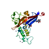 1aolS S: Starting model for refinement |
|---|---|
| Similar structure data |


-Links
 PDBj
PDBj- Assembly
| Deposited unit | 
| |||||||||
|---|---|---|---|---|---|---|---|---|---|---|
| 1 | 
| |||||||||
| 2 | 
| |||||||||
| 3 | 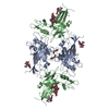
| |||||||||
| 4 | 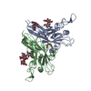
| |||||||||
| Unit cell |
| |||||||||
| Components on special symmetry positions |
|
-Components
| #1: Protein | Mass: 23410.158 Da / Num. of mol.: 2 Source method: isolated from a genetically manipulated source Source: (gene. exp.) Feline leukemia virus / Genus: Gammaretrovirus / Strain: strain B/lambda-B1 / Gene: ENV / Plasmid: PVL1393 / Production host:   Spodoptera frugiperda (fall armyworm) / Strain (production host): SF9 AND HI5 / References: GenBank: 323911, UniProt: P11261*PLUS Spodoptera frugiperda (fall armyworm) / Strain (production host): SF9 AND HI5 / References: GenBank: 323911, UniProt: P11261*PLUS#2: Polysaccharide | Source method: isolated from a genetically manipulated source #3: Polysaccharide | 2-acetamido-2-deoxy-alpha-D-glucopyranose-(1-2)-beta-D-mannopyranose-(1-6)-alpha-D-mannopyranose-(1- ...2-acetamido-2-deoxy-alpha-D-glucopyranose-(1-2)-beta-D-mannopyranose-(1-6)-alpha-D-mannopyranose-(1-4)-2-acetamido-2-deoxy-beta-D-glucopyranose-(1-4)-2-acetamido-2-deoxy-beta-D-glucopyranose | Source method: isolated from a genetically manipulated source #4: Chemical | ChemComp-TOE / |   Mass: 164.200 Da / Num. of mol.: 1 / Source method: obtained synthetically / Formula: C7H16O4 Mass: 164.200 Da / Num. of mol.: 1 / Source method: obtained synthetically / Formula: C7H16O4#5: Water | ChemComp-HOH / |  Mass: 18.015 Da / Num. of mol.: 178 / Source method: isolated from a natural source / Formula: H2O Mass: 18.015 Da / Num. of mol.: 178 / Source method: isolated from a natural source / Formula: H2OHas protein modification | Y | |
|---|
-Experimental details
-Experiment
| Experiment | Method: X-RAY DIFFRACTION / Number of used crystals: 1 |
|---|
- Sample preparation
| Crystal | Density Matthews: 3.11 Å3/Da / Density % sol: 60.46 % | ||||||||||||||||||||||||
|---|---|---|---|---|---|---|---|---|---|---|---|---|---|---|---|---|---|---|---|---|---|---|---|---|---|
| Crystal grow | Temperature: 293 K / Method: vapor diffusion, hanging drop / pH: 5.6 Details: PEG4000, ammonium acetate, sodium citrate, pH 5.6, VAPOR DIFFUSION, HANGING DROP, temperature 293K | ||||||||||||||||||||||||
| Crystal grow | *PLUS | ||||||||||||||||||||||||
| Components of the solutions | *PLUS
|
-Data collection
| Diffraction | Mean temperature: 100 K |
|---|---|
| Diffraction source | Source: SYNCHROTRON / Site: ESRF  / Beamline: ID14-1 / Wavelength: 0.934 Å / Beamline: ID14-1 / Wavelength: 0.934 Å |
| Detector | Type: ADSC QUANTUM 4 / Detector: CCD / Date: Jun 4, 2001 |
| Radiation | Protocol: SINGLE WAVELENGTH / Monochromatic (M) / Laue (L): M / Scattering type: x-ray |
| Radiation wavelength | Wavelength: 0.934 Å / Relative weight: 1 |
| Reflection | Resolution: 2.5→20 Å / Num. all: 21072 / Num. obs: 21010 / % possible obs: 99.7 % / Redundancy: 9 % / Rsym value: 0.061 / Net I/σ(I): 25.4 |
| Reflection shell | Resolution: 2.5→2.59 Å / Mean I/σ(I) obs: 9.4 / Rsym value: 0.192 / % possible all: 100 |
| Reflection | *PLUS Lowest resolution: 20 Å / Num. obs: 21072 / Rmerge(I) obs: 0.061 |
| Reflection shell | *PLUS Highest resolution: 2.5 Å / % possible obs: 100 % / Rmerge(I) obs: 0.192 |
- Processing
| Software |
| ||||||||||||||||||||
|---|---|---|---|---|---|---|---|---|---|---|---|---|---|---|---|---|---|---|---|---|---|
| Refinement | Method to determine structure: MOLECULAR REPLACEMENT Starting model: PDB ENTRY 1AOL Resolution: 2.5→20 Å / Cross valid method: THROUGHOUT / σ(F): 0 / Stereochemistry target values: Engh & Huber
| ||||||||||||||||||||
| Refinement step | Cycle: LAST / Resolution: 2.5→20 Å
| ||||||||||||||||||||
| Refine LS restraints |
| ||||||||||||||||||||
| Refinement | *PLUS Lowest resolution: 20 Å / % reflection Rfree: 7 % | ||||||||||||||||||||
| Solvent computation | *PLUS | ||||||||||||||||||||
| Displacement parameters | *PLUS |
