 Movie
Movie Controller

[English] 日本語
 Yorodumi
- PDB-4r29: Crystal structure of bacterial cysteine methyltransferase effecto... -
+ Open data

- Basic information
| Entry | Database: PDB / ID: 4r29 | ||||||
|---|---|---|---|---|---|---|---|
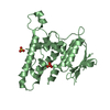
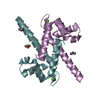
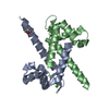
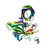
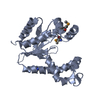
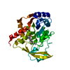
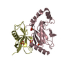
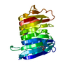
| Title | Crystal structure of bacterial cysteine methyltransferase effector NleE | ||||||
 Components Components | Uncharacterized protein | ||||||
 Keywords Keywords | TRANSFERASE / Rossmann-like Fold / NF-kappaB inhibition / TAB2 and TAB3 | ||||||
| Function / homology |  Function and homology information Function and homology informationTransferases; Transferring one-carbon groups; Methyltransferases / methyltransferase activity / toxin activity / methylation / host cell nucleus / extracellular region Similarity search - Function | ||||||
| Biological species |  | ||||||
| Method |  X-RAY DIFFRACTION / SYNCHROTRON / SAD / Resolution: 2.31 Å X-RAY DIFFRACTION / SYNCHROTRON / SAD / Resolution: 2.31 Å | ||||||
 Authors Authors | Yao, Q. / Chen, J. / Hu, L. / Zhang, L. / Shao, F. | ||||||
 Citation Citation | Journal: Plos Pathog. / Year: 2014 Title: Structure and Specificity of the Bacterial Cysteine Methyltransferase Effector NleE Suggests a Novel Substrate in Human DNA Repair Pathway. Authors: Yao, Q. / Zhang, L. / Wan, X. / Chen, J. / Hu, L. / Ding, X. / Li, L. / Karar, J. / Peng, H. / Chen, S. / Huang, N. / Rauscher, F.J. / Shao, F. | ||||||
| History |
|
- Structure visualization
| Structure viewer | Molecule: MolmilJmol/JSmol |
|---|
- Downloads & links
-Download
| PDBx/mmCIF format | 4r29.cif.gz | 181.1 KB | Display | PDBx/mmCIF format |
|---|---|---|---|---|
| PDB format | pdb4r29.ent.gz | 145.1 KB | Display | PDB format |
| PDBx/mmJSON format | 4r29.json.gz | Tree view | PDBx/mmJSON format | |
| Others |  Other downloads Other downloads |
-Validation report
| Arichive directory | https://data.pdbj.org/pub/pdb/validation_reports/r2/4r29ftp://data.pdbj.org/pub/pdb/validation_reports/r2/4r29 | HTTPS FTP |
|---|
-Related structure data
| Similar structure data |
|---|


-Links
 PDBj
PDBj
- Assembly
| Deposited unit | 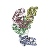
| ||||||||
|---|---|---|---|---|---|---|---|---|---|
| 1 | 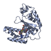
| ||||||||
| 2 | 
| ||||||||
| 3 | 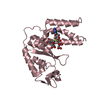
| ||||||||
| 4 | 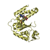
| ||||||||
| Unit cell |
|
-Components
| #1: Protein | Mass: 26029.771 Da / Num. of mol.: 4 / Mutation: E181A Source method: isolated from a genetically manipulated source Source: (gene. exp.) #2: Chemical | ChemComp-SAM /   Mass: 398.437 Da / Num. of mol.: 4 / Source method: obtained synthetically / Formula: C15H22N6O5S Mass: 398.437 Da / Num. of mol.: 4 / Source method: obtained synthetically / Formula: C15H22N6O5S#3: Chemical |   Mass: 92.094 Da / Num. of mol.: 3 / Source method: obtained synthetically / Formula: C3H8O3 Mass: 92.094 Da / Num. of mol.: 3 / Source method: obtained synthetically / Formula: C3H8O3#4: Chemical |   Mass: 192.124 Da / Num. of mol.: 2 / Source method: obtained synthetically / Formula: C6H8O7 Mass: 192.124 Da / Num. of mol.: 2 / Source method: obtained synthetically / Formula: C6H8O7#5: Water | ChemComp-HOH / |  Mass: 18.015 Da / Num. of mol.: 295 / Source method: isolated from a natural source / Formula: H2O Mass: 18.015 Da / Num. of mol.: 295 / Source method: isolated from a natural source / Formula: H2O |
|---|
-Experimental details
-Experiment
| Experiment | Method: X-RAY DIFFRACTION / Number of used crystals: 1 |
|---|
- Sample preparation
| Crystal | Density Matthews: 2.34 Å3/Da / Density % sol: 47.46 % |
|---|
-Data collection
| Diffraction | Mean temperature: 200 K |
|---|---|
| Diffraction source | Source: SYNCHROTRON / Site: SSRF  / Beamline: BL17U / Wavelength: 0.9791 Å / Beamline: BL17U / Wavelength: 0.9791 Å |
| Detector | Type: ADSC QUANTUM 315r / Detector: CCD / Date: May 20, 2011 |
| Radiation | Protocol: SINGLE WAVELENGTH / Monochromatic (M) / Laue (L): M / Scattering type: x-ray |
| Radiation wavelength | Wavelength: 0.9791 Å / Relative weight: 1 |
| Reflection | Resolution: 2.3→20.02 Å / Num. all: 43485 / Num. obs: 42616 / % possible obs: 99.8 % / Observed criterion σ(F): 261.7 / Observed criterion σ(I): 57.3 / Redundancy: 3.7 % |
| Reflection shell | Resolution: 2.3→2.34 Å / % possible all: 98.4 |
- Processing
| Software |
| ||||||||||||||||||||||||||||||||||||||||||||||||||||||||||||||||||
|---|---|---|---|---|---|---|---|---|---|---|---|---|---|---|---|---|---|---|---|---|---|---|---|---|---|---|---|---|---|---|---|---|---|---|---|---|---|---|---|---|---|---|---|---|---|---|---|---|---|---|---|---|---|---|---|---|---|---|---|---|---|---|---|---|---|---|---|
| Refinement | Method to determine structure: SAD / Resolution: 2.31→20.02 Å / SU ML: 0.26 / σ(F): 0.04 / Phase error: 23.85 / Stereochemistry target values: MLHL
| ||||||||||||||||||||||||||||||||||||||||||||||||||||||||||||||||||
| Solvent computation | Shrinkage radii: 0.9 Å / VDW probe radii: 1.11 Å / Solvent model: FLAT BULK SOLVENT MODEL / Bsol: 46.751 Å2 / ksol: 0.4 e/Å3 | ||||||||||||||||||||||||||||||||||||||||||||||||||||||||||||||||||
| Displacement parameters |
| ||||||||||||||||||||||||||||||||||||||||||||||||||||||||||||||||||
| Refinement step | Cycle: LAST / Resolution: 2.31→20.02 Å
| ||||||||||||||||||||||||||||||||||||||||||||||||||||||||||||||||||
| Refine LS restraints |
| ||||||||||||||||||||||||||||||||||||||||||||||||||||||||||||||||||
| LS refinement shell | Refine-ID: X-RAY DIFFRACTION / Total num. of bins used: 10
|
