- PDB-3k0y: Crystal structure of Putative TOXIN related protein (YP_001303978... -
+
Open data
ID or keywords:
Loading...
-
Basic information
Entry
Database: PDB / ID: 3k0y
Title
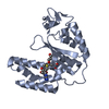
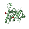
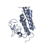
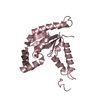
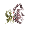
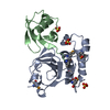
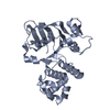
Crystal structure of Putative TOXIN related protein (YP_001303978.1) from Parabacteroides distasonis ATCC 8503 at 2.16 A resolution
Components
Putative TOXIN related protein
Keywords
TOXIN / Putative TOXIN related protein / Structural Genomics / Joint Center for Structural Genomics / JCSG / Protein Structure Initiative / PSI-2 / unknown function
Mass: 18.015 Da / Num. of mol.: 56 / Source method: isolated from a natural source / Formula: H2O
Has protein modification
Y
Sequence details
THIS CONSTRUCT (RESIDUE 26-253) WAS EXPRESSED WITH A PURIFICATION TAG MGSDKIHHHHHHENLYFQG. THE TAG ...THIS CONSTRUCT (RESIDUE 26-253) WAS EXPRESSED WITH A PURIFICATION TAG MGSDKIHHHHHHENLYFQG. THE TAG WAS REMOVED WITH TEV PROTEASE LEAVING ONLY A GLYCINE (0) FOLLOWED BY THE TARGET SEQUENCE.
-
Experimental details
-
Experiment
Experiment
Method: X-RAY DIFFRACTION / Number of used crystals: 2
Resolution: 2.16→39.559 Å / Num. obs: 16892 / % possible obs: 99.4 % / Observed criterion σ(I): -3 / Biso Wilson estimate: 52.236 Å2 / Rmerge(I) obs: 0.032 / Net I/σ(I): 19.28
Reflection shell
Resolution (Å)
Rmerge(I) obs
Mean I/σ(I) obs
Num. measured obs
Num. unique obs
Diffraction-ID
% possible all
2.16-2.24
0.688
1.8
9460
3289
1,2
99.5
2.24-2.33
0.466
2.7
8862
3088
1,2
99.3
2.33-2.43
0.318
3.6
8586
2986
1,2
99.7
2.43-2.56
0.241
4.9
9434
3260
1,2
99.6
2.56-2.72
0.152
7.5
8968
3097
1,2
99.5
2.72-2.93
0.087
12.6
9163
3157
1,2
99.4
2.93-3.22
0.048
20.8
9090
3138
1,2
99.6
3.22-3.69
0.025
35.1
9233
3172
1,2
99.4
3.69-4.64
0.018
48.7
9169
3152
1,2
99.5
4.64-39.559
0.017
54.9
9091
3156
1,2
98.3
-
Phasing
Phasing
Method: MAD
-
Processing
Software
Name
Version
Classification
NB
REFMAC
5.5.0053
refinement
PHENIX
refinement
MolProbity
3beta29
modelbuilding
XSCALE
datascaling
PDB_EXTRACT
3.006
dataextraction
XDS
datareduction
SHARP
phasing
autoSHARP
phasing
Refinement
Method to determine structure: MAD / Resolution: 2.16→39.559 Å / Cor.coef. Fo:Fc: 0.954 / Cor.coef. Fo:Fc free: 0.937 / Occupancy max: 1 / Occupancy min: 0.5 / SU B: 10.948 / SU ML: 0.129 / TLS residual ADP flag: LIKELY RESIDUAL / Cross valid method: THROUGHOUT / σ(F): 0 / ESU R: 0.189 / ESU R Free: 0.167 / Stereochemistry target values: MAXIMUM LIKELIHOOD Details: 1. HYDROGENS HAVE BEEN ADDED IN THE RIDING POSITIONS. 2. ATOM RECORDS CONTAIN RESIDUAL B FACTORS ONLY. 3. A MET-INHIBITION PROTOCOL WAS USED FOR SELENOMETHIONINE INCORPORATION DURING PROTEIN ...Details: 1. HYDROGENS HAVE BEEN ADDED IN THE RIDING POSITIONS. 2. ATOM RECORDS CONTAIN RESIDUAL B FACTORS ONLY. 3. A MET-INHIBITION PROTOCOL WAS USED FOR SELENOMETHIONINE INCORPORATION DURING PROTEIN EXPRESSION. THE OCCUPANCY OF THE SE ATOMS IN THE MSE RESIDUES WAS REDUCED TO 0.75 FOR THE REDUCED SCATTERING POWER DUE TO PARTIAL S-MET INCORPORATION. 4. PEG-400 (2PE) FRAGMENTS FROM THE CRYSTALLIZATION SOLUTION ARE MODELED.
Rfactor
Num. reflection
% reflection
Selection details
Rfree
0.235
853
5.1 %
RANDOM
Rwork
0.203
-
-
-
obs
0.205
16861
99.85 %
-
Solvent computation
Ion probe radii: 0.8 Å / Shrinkage radii: 0.8 Å / VDW probe radii: 1.4 Å / Solvent model: BABINET MODEL WITH MASK
In the structure databanks used in Yorodumi, some data are registered as the other names, "COVID-19 virus" and "2019-nCoV". Here are the details of the virus and the list of structure data.
Jan 31, 2019. EMDB accession codes are about to change! (news from PDBe EMDB page)
EMDB accession codes are about to change! (news from PDBe EMDB page)
The allocation of 4 digits for EMDB accession codes will soon come to an end. Whilst these codes will remain in use, new EMDB accession codes will include an additional digit and will expand incrementally as the available range of codes is exhausted. The current 4-digit format prefixed with “EMD-” (i.e. EMD-XXXX) will advance to a 5-digit format (i.e. EMD-XXXXX), and so on. It is currently estimated that the 4-digit codes will be depleted around Spring 2019, at which point the 5-digit format will come into force.
The EM Navigator/Yorodumi systems omit the EMD- prefix.
Related info.:Q: What is EMD? / ID/Accession-code notation in Yorodumi/EM Navigator
Yorodumi is a browser for structure data from EMDB, PDB, SASBDB, etc.
This page is also the successor to EM Navigator detail page, and also detail information page/front-end page for Omokage search.
The word "yorodu" (or yorozu) is an old Japanese word meaning "ten thousand". "mi" (miru) is to see.
Related info.:EMDB / PDB / SASBDB / Comparison of 3 databanks / Yorodumi Search / Aug 31, 2016. New EM Navigator & Yorodumi / Yorodumi Papers / Jmol/JSmol / Function and homology information / Changes in new EM Navigator and Yorodumi
 Movie
Movie Controller

 Yorodumi
 Open data
 Basic information
 Components
 Keywords
 Function and homology information
 Parabacteroides distasonis ATCC 8503 (bacteria)
 X-RAY DIFFRACTION /
  Authors
 Citation
 Structure visualization
 Downloads & links
 Other downloads
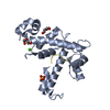


 PDBj
PDBj Assembly

 Mass: 414.488 Da / Num. of mol.: 2 / Source method: obtained synthetically / Formula: C18H38O10 / Comment: precipitant*YM
 Mass: 18.015 Da / Num. of mol.: 56 / Source method: isolated from a natural source / Formula: H2O
 Sample preparation
 Processing