 Movie
Movie Controller

[English] 日本語
 Yorodumi
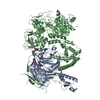
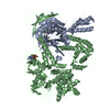
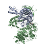
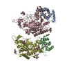
- PDB-1l1o: Structure of the human Replication Protein A (RPA) trimerization core -
+ Open data

- Basic information
| Entry | Database: PDB / ID: 1l1o | ||||||
|---|---|---|---|---|---|---|---|
| Title | Structure of the human Replication Protein A (RPA) trimerization core | ||||||
 Components Components |
| ||||||
 Keywords Keywords | DNA BINDING PROTEIN / eukaryotic SSB / ssDNA binding protein / OB-fold | ||||||
| Function / homology |  Function and homology information Function and homology informationprotein localization to chromosome / DNA replication factor A complex / single-stranded telomeric DNA binding / Removal of the Flap Intermediate / lateral element / G-rich strand telomeric DNA binding / protein localization to site of double-strand break / Mismatch repair (MMR) directed by MSH2:MSH3 (MutSbeta) / Mismatch repair (MMR) directed by MSH2:MSH6 (MutSalpha) / regulation of DNA damage checkpoint ...protein localization to chromosome / DNA replication factor A complex / single-stranded telomeric DNA binding / Removal of the Flap Intermediate / lateral element / G-rich strand telomeric DNA binding / protein localization to site of double-strand break / Mismatch repair (MMR) directed by MSH2:MSH3 (MutSbeta) / Mismatch repair (MMR) directed by MSH2:MSH6 (MutSalpha) / regulation of DNA damage checkpoint / chromatin-protein adaptor activity / Removal of the Flap Intermediate from the C-strand / regulation of double-strand break repair via homologous recombination / HDR through Single Strand Annealing (SSA) / telomeric repeat DNA binding / Impaired BRCA2 binding to RAD51 / hemopoiesis / PCNA-Dependent Long Patch Base Excision Repair / Activation of the pre-replicative complex / Regulation of HSF1-mediated heat shock response / Presynaptic phase of homologous DNA pairing and strand exchange / HSF1 activation / Activation of ATR in response to replication stress / telomere maintenance via telomerase / mismatch repair / SUMOylation of DNA damage response and repair proteins / site of DNA damage / mitotic G1 DNA damage checkpoint signaling / regulation of mitotic cell cycle / homeostasis of number of cells within a tissue / telomere maintenance / Translesion synthesis by REV1 / Translesion synthesis by POLK / male germ cell nucleus / Translesion synthesis by POLI / Gap-filling DNA repair synthesis and ligation in GG-NER / meiotic cell cycle / nucleotide-excision repair / Fanconi Anemia Pathway / Termination of translesion DNA synthesis / Translesion Synthesis by POLH / Recognition of DNA damage by PCNA-containing replication complex / base-excision repair / PML body / G2/M DNA damage checkpoint / DNA-templated DNA replication / double-strand break repair via homologous recombination / Meiotic recombination / HDR through Homologous Recombination (HRR) / Dual Incision in GG-NER / Formation of Incision Complex in GG-NER / Dual incision in TC-NER / Gap-filling DNA repair synthesis and ligation in TC-NER / regulation of cell population proliferation / single-stranded DNA binding / site of double-strand break / Processing of DNA double-strand break ends / DNA recombination / protein phosphatase binding / Regulation of TP53 Activity through Phosphorylation / in utero embryonic development / damaged DNA binding / DNA replication / chromosome, telomeric region / nuclear body / DNA repair / positive regulation of cell population proliferation / chromatin binding / DNA damage response / ubiquitin protein ligase binding / chromatin / enzyme binding / zinc ion binding / nucleoplasm / nucleus Similarity search - Function | ||||||
| Biological species |  Homo sapiens (human) Homo sapiens (human) | ||||||
| Method |  X-RAY DIFFRACTION / SYNCHROTRON / MAD / Resolution: 2.8 Å X-RAY DIFFRACTION / SYNCHROTRON / MAD / Resolution: 2.8 Å | ||||||
 Authors Authors | Bochkareva, E.V. / Korolev, S. / Lees-Miller, S.P. / Bochkarev, A. | ||||||
 Citation Citation | Journal: EMBO J. / Year: 2002 Title: Structure of the RPA trimerization core and its role in the multistep DNA-binding mechanism of RPA. Authors: Bochkareva, E. / Korolev, S. / Lees-Miller, S.P. / Bochkarev, A. #1: Journal: J.Biol.Chem. / Year: 2000Title: The role for zinc in replication protein A. Authors: Bochkareva, E. / Korolev, S. / Bochkarev, A. | ||||||
| History |
| ||||||
| Remark 650 | HELIX AUTHOR PROVIDED SECONDARY STRUCTURE. | ||||||
| Remark 700 | SHEET AUTHOR PROVIDED SHEET RECORDS. |
- Structure visualization
| Structure viewer | Molecule: MolmilJmol/JSmol |
|---|
- Downloads & links
-Download
| PDBx/mmCIF format | 1l1o.cif.gz | 174.6 KB | Display | PDBx/mmCIF format |
|---|---|---|---|---|
| PDB format | pdb1l1o.ent.gz | 138.7 KB | Display | PDB format |
| PDBx/mmJSON format | 1l1o.json.gz | Tree view | PDBx/mmJSON format | |
| Others |  Other downloads Other downloads |
-Validation report
| Arichive directory | https://data.pdbj.org/pub/pdb/validation_reports/l1/1l1oftp://data.pdbj.org/pub/pdb/validation_reports/l1/1l1o | HTTPS FTP |
|---|
-Related structure data
| Related structure data | |
|---|---|
| Similar structure data |


-Links
 PDBj
PDBj


- Assembly
| Deposited unit | 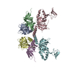
| ||||||||
|---|---|---|---|---|---|---|---|---|---|
| 1 |
| ||||||||
| Unit cell |
| ||||||||
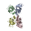
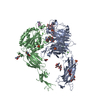
| Details | RPA is a heterotrimer made of RPA70, RPA32, and RPA14 subunits |
-Components
| #1: Protein | Mass: 13583.714 Da / Num. of mol.: 2 / Fragment: RPA14 Source method: isolated from a genetically manipulated source Source: (gene. exp.) Homo sapiens (human) / Gene: RFA3_HUMAN / Plasmid: pET15B 3-core / Species (production host): Escherichia coli / Production host:  #2: Protein | Mass: 14361.513 Da / Num. of mol.: 2 / Fragment: RPA32 central domain (residues 44-171) Source method: isolated from a genetically manipulated source Source: (gene. exp.) Homo sapiens (human) / Gene: RFA2_HUMAN / Plasmid: pET15B 3-core / Species (production host): Escherichia coli / Production host: #3: Protein | Mass: 21145.850 Da / Num. of mol.: 2 / Fragment: RPA70 C-terminal domain (residues 436-616) Source method: isolated from a genetically manipulated source Source: (gene. exp.) Homo sapiens (human) / Gene: RFA1_HUMAN / Plasmid: pET15B 3-core / Species (production host): Escherichia coli / Production host: #4: Chemical |   Mass: 65.409 Da / Num. of mol.: 2 / Source method: obtained synthetically / Formula: Zn Mass: 65.409 Da / Num. of mol.: 2 / Source method: obtained synthetically / Formula: Zn |
|---|
-Experimental details
-Experiment
| Experiment | Method: X-RAY DIFFRACTION / Number of used crystals: 1 |
|---|
- Sample preparation
| Crystal | Density Matthews: 3.93 Å3/Da / Density % sol: 68.69 % | ||||||||||||||||||||||||
|---|---|---|---|---|---|---|---|---|---|---|---|---|---|---|---|---|---|---|---|---|---|---|---|---|---|
| Crystal grow | Temperature: 300 K / Method: vapor diffusion, sitting drop / pH: 7.8 Details: 0.1 M Hepes, 9% glycerol, and 1.6 M ammonium sulfate, pH 7.8, VAPOR DIFFUSION, SITTING DROP, temperature 300K | ||||||||||||||||||||||||
| Crystal grow | *PLUS Details: Bochkareva, E., (2000) J.Biol.Chem., 275, 27332. | ||||||||||||||||||||||||
| Components of the solutions | *PLUS
|
-Data collection
| Diffraction |
| ||||||||||||||||||
|---|---|---|---|---|---|---|---|---|---|---|---|---|---|---|---|---|---|---|---|
| Diffraction source |
| ||||||||||||||||||
| Detector |
| ||||||||||||||||||
| Radiation | Protocol: MAD / Monochromatic (M) / Laue (L): M / Scattering type: x-ray | ||||||||||||||||||
| Radiation wavelength |
| ||||||||||||||||||
| Reflection | Resolution: 2.7→20 Å / Num. obs: 43573 / % possible obs: 99.2 % / Observed criterion σ(I): -3 / Redundancy: 5.4 % / Biso Wilson estimate: 68.5 Å2 / Rsym value: 0.062 / Net I/σ(I): 2.7 | ||||||||||||||||||
| Reflection shell | Resolution: 2.7→2.8 Å / Mean I/σ(I) obs: 2.7 / Num. unique all: 4302 / Rsym value: 0.532 / % possible all: 99.7 | ||||||||||||||||||
| Reflection | *PLUS Num. measured all: 233991 / Rmerge(I) obs: 0.062 | ||||||||||||||||||
| Reflection shell | *PLUS % possible obs: 99.7 % / Rmerge(I) obs: 0.532 |

- Processing
| Software |
| ||||||||||||||||||||||||||||||||||||
|---|---|---|---|---|---|---|---|---|---|---|---|---|---|---|---|---|---|---|---|---|---|---|---|---|---|---|---|---|---|---|---|---|---|---|---|---|---|
| Refinement | Method to determine structure: MAD / Resolution: 2.8→19.92 Å / Rfactor Rfree error: 0.005 / Data cutoff high absF: 584448.25 / Data cutoff high rms absF: 584448.25 / Data cutoff low absF: 0 / Isotropic thermal model: RESTRAINED / Cross valid method: THROUGHOUT / σ(F): 2 / Stereochemistry target values: Engh & Huber
| ||||||||||||||||||||||||||||||||||||
| Solvent computation | Solvent model: FLAT MODEL / Bsol: 39.918 Å2 / ksol: 0.323875 e/Å3 | ||||||||||||||||||||||||||||||||||||
| Displacement parameters | Biso mean: 59.9 Å2
| ||||||||||||||||||||||||||||||||||||
| Refine analyze |
| ||||||||||||||||||||||||||||||||||||
| Refinement step | Cycle: LAST / Resolution: 2.8→19.92 Å
| ||||||||||||||||||||||||||||||||||||
| Refine LS restraints |
| ||||||||||||||||||||||||||||||||||||
| LS refinement shell | Resolution: 2.8→2.97 Å / Rfactor Rfree error: 0.017 / Total num. of bins used: 6
| ||||||||||||||||||||||||||||||||||||
| Xplor file |
| ||||||||||||||||||||||||||||||||||||
| Refinement | *PLUS % reflection Rfree: 9.4 % / Rfactor all: 0.245 / Rfactor obs: 0.236 / Rfactor Rfree: 0.283 / Rfactor Rwork: 0.236 | ||||||||||||||||||||||||||||||||||||
| Solvent computation | *PLUS | ||||||||||||||||||||||||||||||||||||
| Displacement parameters | *PLUS | ||||||||||||||||||||||||||||||||||||
| Refine LS restraints | *PLUS
| ||||||||||||||||||||||||||||||||||||
| LS refinement shell | *PLUS Rfactor Rfree: 0.373 / Rfactor Rwork: 0.336 / Rfactor obs: 0.336 |
