 Movie
Movie Controller

[English] 日本語
 Yorodumi
- PDB-1fgu: SSDNA-BINDING DOMAIN OF THE LARGE SUBUNIT OF REPLICATION PROTEIN A -
+ Open data

- Basic information
| Entry | Database: PDB / ID: 1fgu | ||||||
|---|---|---|---|---|---|---|---|
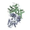
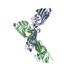
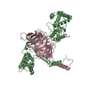
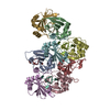
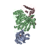
| Title | SSDNA-BINDING DOMAIN OF THE LARGE SUBUNIT OF REPLICATION PROTEIN A | ||||||
 Components Components | REPLICATION PROTEIN A 70 KDA DNA-BINDING SUBUNIT | ||||||
 Keywords Keywords | REPLICATION / OB-fold / ssDNA-binding protein | ||||||
| Function / homology |  Function and homology information Function and homology informationprotein localization to chromosome / DNA replication factor A complex / single-stranded telomeric DNA binding / Removal of the Flap Intermediate / lateral element / G-rich strand telomeric DNA binding / protein localization to site of double-strand break / Mismatch repair (MMR) directed by MSH2:MSH3 (MutSbeta) / Mismatch repair (MMR) directed by MSH2:MSH6 (MutSalpha) / chromatin-protein adaptor activity ...protein localization to chromosome / DNA replication factor A complex / single-stranded telomeric DNA binding / Removal of the Flap Intermediate / lateral element / G-rich strand telomeric DNA binding / protein localization to site of double-strand break / Mismatch repair (MMR) directed by MSH2:MSH3 (MutSbeta) / Mismatch repair (MMR) directed by MSH2:MSH6 (MutSalpha) / chromatin-protein adaptor activity / Removal of the Flap Intermediate from the C-strand / HDR through Single Strand Annealing (SSA) / Impaired BRCA2 binding to RAD51 / hemopoiesis / PCNA-Dependent Long Patch Base Excision Repair / Activation of the pre-replicative complex / Regulation of HSF1-mediated heat shock response / Presynaptic phase of homologous DNA pairing and strand exchange / HSF1 activation / Activation of ATR in response to replication stress / telomere maintenance via telomerase / mismatch repair / SUMOylation of DNA damage response and repair proteins / site of DNA damage / homeostasis of number of cells within a tissue / telomere maintenance / Translesion synthesis by REV1 / Translesion synthesis by POLK / male germ cell nucleus / Translesion synthesis by POLI / Gap-filling DNA repair synthesis and ligation in GG-NER / meiotic cell cycle / nucleotide-excision repair / Fanconi Anemia Pathway / Termination of translesion DNA synthesis / Translesion Synthesis by POLH / Recognition of DNA damage by PCNA-containing replication complex / base-excision repair / PML body / G2/M DNA damage checkpoint / DNA-templated DNA replication / double-strand break repair via homologous recombination / Meiotic recombination / HDR through Homologous Recombination (HRR) / Dual Incision in GG-NER / Formation of Incision Complex in GG-NER / Dual incision in TC-NER / Gap-filling DNA repair synthesis and ligation in TC-NER / single-stranded DNA binding / site of double-strand break / Processing of DNA double-strand break ends / DNA recombination / Regulation of TP53 Activity through Phosphorylation / in utero embryonic development / damaged DNA binding / DNA replication / chromosome, telomeric region / DNA repair / positive regulation of cell population proliferation / chromatin binding / DNA damage response / zinc ion binding / nucleoplasm / nucleus Similarity search - Function | ||||||
| Biological species |  Homo sapiens (human) Homo sapiens (human) | ||||||
| Method |  X-RAY DIFFRACTION / SYNCHROTRON / MAD / Resolution: 2.5 Å X-RAY DIFFRACTION / SYNCHROTRON / MAD / Resolution: 2.5 Å | ||||||
 Authors Authors | Bochkareva, E. / Belegu, V. / Korolev, S. / Bochkarev, A. | ||||||
 Citation Citation | Journal: EMBO J. / Year: 2001 Title: Structure of the major single-stranded DNA-binding domain of replication protein A suggests a dynamic mechanism for DNA binding. Authors: Bochkareva, E. / Belegu, V. / Korolev, S. / Bochkarev, A. #1: Journal: Nature / Year: 1997Title: Structure of the Single-stranded-DNA-binding Domain of Replication Protein A Bound to DNA Authors: Bochkarev, A. / Pfuetzner, R.A. / Edwards, A.M. / Frappier, L. #2: Journal: J.Biol.Chem. / Year: 1997Title: Replication Protein A: Characterization and Crystallization of the DNA-binding Domain Authors: Pfuetzner, R.A. / Bochkarev, A. / Frappier, L. / Edwards, A.M. | ||||||
| History |
|
- Structure visualization
| Structure viewer | Molecule: MolmilJmol/JSmol |
|---|
- Downloads & links
-Download
| PDBx/mmCIF format | 1fgu.cif.gz | 104.6 KB | Display | PDBx/mmCIF format |
|---|---|---|---|---|
| PDB format | pdb1fgu.ent.gz | 82.2 KB | Display | PDB format |
| PDBx/mmJSON format | 1fgu.json.gz | Tree view | PDBx/mmJSON format | |
| Others |  Other downloads Other downloads |
-Validation report
| Arichive directory | https://data.pdbj.org/pub/pdb/validation_reports/fg/1fguftp://data.pdbj.org/pub/pdb/validation_reports/fg/1fgu | HTTPS FTP |
|---|
-Related structure data
| Related structure data | |
|---|---|
| Similar structure data |


-Links
 PDBj
PDBj


- Assembly
| Deposited unit | 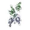
| ||||||||
|---|---|---|---|---|---|---|---|---|---|
| 1 |
| ||||||||
| Unit cell |
| ||||||||
| Details | The biological assembly is a trimer of 70, 32, and 14 kDa subunit THE BIOLOGICAL ASSEMBLY IS A TRIMER OF 70, 32, and 14 KDA SUBUNITS |
-Components
| #1: Protein | Mass: 28109.641 Da / Num. of mol.: 2 / Fragment: CENTRAL DOMAIN Source method: isolated from a genetically manipulated source Source: (gene. exp.) Homo sapiens (human) / Plasmid details: BL21(DE3) / Production host:  #2: Water | ChemComp-HOH / |  Mass: 18.015 Da / Num. of mol.: 32 / Source method: isolated from a natural source / Formula: H2O Mass: 18.015 Da / Num. of mol.: 32 / Source method: isolated from a natural source / Formula: H2O |
|---|
-Experimental details
-Experiment
| Experiment | Method: X-RAY DIFFRACTION / Number of used crystals: 1 |
|---|
- Sample preparation
| Crystal | Density Matthews: 2.86 Å3/Da / Density % sol: 57 % | ||||||||||||||||||||||||||||||||||||||||||||||||
|---|---|---|---|---|---|---|---|---|---|---|---|---|---|---|---|---|---|---|---|---|---|---|---|---|---|---|---|---|---|---|---|---|---|---|---|---|---|---|---|---|---|---|---|---|---|---|---|---|---|
| Crystal grow | Temperature: 298 K / Method: vapor diffusion, sitting drop / pH: 8.5 Details: 0.1 M K,NaH2PO4 0.1 M Tris 3.8 M NaCl, pH 8.5, VAPOR DIFFUSION, SITTING DROP, temperature 298K | ||||||||||||||||||||||||||||||||||||||||||||||||
| Crystal grow | *PLUS pH: 7 | ||||||||||||||||||||||||||||||||||||||||||||||||
| Components of the solutions | *PLUS
|
-Data collection
| Diffraction | Mean temperature: 100 K |
|---|---|
| Diffraction source | Source: SYNCHROTRON / Site: APS  / Beamline: 19-ID / Wavelength: 1.033 / Beamline: 19-ID / Wavelength: 1.033 |
| Detector | Type: SBC-2 / Detector: CCD / Date: Jan 27, 2000 |
| Radiation | Protocol: MAD / Monochromatic (M) / Laue (L): M / Scattering type: x-ray |
| Radiation wavelength | Wavelength: 1.033 Å / Relative weight: 1 |
| Reflection | Resolution: 2.5→30 Å / Num. all: 247836 / Num. obs: 22963 / % possible obs: 98.6 % / Observed criterion σ(F): 0 / Observed criterion σ(I): -3 / Redundancy: 10.8 % / Biso Wilson estimate: 57.6 Å2 / Rmerge(I) obs: 0.052 / Net I/σ(I): 37.1 |
| Reflection shell | Resolution: 2.49→2.58 Å / Rmerge(I) obs: 0.19 / Mean I/σ(I) obs: 4.6 / Num. unique all: 2191 / % possible all: 96 |
| Reflection | *PLUS Num. measured all: 247836 |
| Reflection shell | *PLUS % possible obs: 96 % / Rmerge(I) obs: 0.19 |
- Processing
| Software |
| ||||||||||||||||||||||||||||||||||||||||||||||||||||||||||||
|---|---|---|---|---|---|---|---|---|---|---|---|---|---|---|---|---|---|---|---|---|---|---|---|---|---|---|---|---|---|---|---|---|---|---|---|---|---|---|---|---|---|---|---|---|---|---|---|---|---|---|---|---|---|---|---|---|---|---|---|---|---|
| Refinement | Method to determine structure: MAD / Resolution: 2.5→20 Å / σ(F): 1.5 / σ(I): 0 / Stereochemistry target values: ENGH & HUBER / Details: CRYSTALS DIFFRACTED ANYSOTROPICALLY
| ||||||||||||||||||||||||||||||||||||||||||||||||||||||||||||
| Solvent computation | Solvent model: CNS DEFAULT BULK SOLVENT / Bsol: 39.1 Å2 / ksol: 0.333 e/Å3 | ||||||||||||||||||||||||||||||||||||||||||||||||||||||||||||
| Displacement parameters | Biso mean: 56.99 Å2
| ||||||||||||||||||||||||||||||||||||||||||||||||||||||||||||
| Refine analyze |
| ||||||||||||||||||||||||||||||||||||||||||||||||||||||||||||
| Refinement step | Cycle: LAST / Resolution: 2.5→20 Å
| ||||||||||||||||||||||||||||||||||||||||||||||||||||||||||||
| Refine LS restraints |
| ||||||||||||||||||||||||||||||||||||||||||||||||||||||||||||
| LS refinement shell | Resolution: 2.5→2.59 Å / Total num. of bins used: 10
| ||||||||||||||||||||||||||||||||||||||||||||||||||||||||||||
| Xplor file |
| ||||||||||||||||||||||||||||||||||||||||||||||||||||||||||||
| Software | *PLUS Name: CNS / Version: 1 / Classification: refinement | ||||||||||||||||||||||||||||||||||||||||||||||||||||||||||||
| Refinement | *PLUS | ||||||||||||||||||||||||||||||||||||||||||||||||||||||||||||
| Solvent computation | *PLUS | ||||||||||||||||||||||||||||||||||||||||||||||||||||||||||||
| Displacement parameters | *PLUS | ||||||||||||||||||||||||||||||||||||||||||||||||||||||||||||
| Refine LS restraints | *PLUS
| ||||||||||||||||||||||||||||||||||||||||||||||||||||||||||||
| LS refinement shell | *PLUS % reflection Rfree: 4.4 % |
