 Movie
Movie Controller

[English] 日本語
 Yorodumi
- PDB-1kw0: Catalytic Domain of Human Phenylalanine Hydroxylase (Fe(II)) in C... -
+ Open data

- Basic information
| Entry | Database: PDB / ID: 1kw0 | ||||||
|---|---|---|---|---|---|---|---|
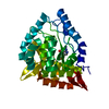
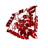
| Title | Catalytic Domain of Human Phenylalanine Hydroxylase (Fe(II)) in Complex with Tetrahydrobiopterin and Thienylalanine | ||||||
 Components Components | Phenylalanine-4-hydroxylase | ||||||
 Keywords Keywords | OXIDOREDUCTASE / Basket-arrangement 13 alpha-helices and 6 beta-strands / Ferrous iron | ||||||
| Function / homology |  Function and homology information Function and homology informationPhenylketonuria / phenylalanine 4-monooxygenase / Phenylalanine metabolism / phenylalanine 4-monooxygenase activity / L-tyrosine biosynthetic process / catecholamine biosynthetic process / L-phenylalanine catabolic process / amino acid biosynthetic process / iron ion binding / cytosol Similarity search - Function | ||||||
| Biological species |  Homo sapiens (human) Homo sapiens (human) | ||||||
| Method |  X-RAY DIFFRACTION / SYNCHROTRON / adopting entry 1PAH phases / Resolution: 2.5 Å X-RAY DIFFRACTION / SYNCHROTRON / adopting entry 1PAH phases / Resolution: 2.5 Å | ||||||
 Authors Authors | Andersen, O.A. / Flatmark, T. / Hough, E. | ||||||
 Citation Citation | Journal: J.Mol.Biol. / Year: 2002 Title: Crystal Structure of the Ternary Complex of the Catalytic Domain of Human Phenylalanine Hydroxylase with Tetrahydrobiopterin and 3-(2-thienyl)-L-alanine, and its Implications for the Mechanism ...Title: Crystal Structure of the Ternary Complex of the Catalytic Domain of Human Phenylalanine Hydroxylase with Tetrahydrobiopterin and 3-(2-thienyl)-L-alanine, and its Implications for the Mechanism of Catalysis and Substrate Activation Authors: Andersen, O.A. / Flatmark, T. / Hough, E. | ||||||
| History |
|
- Structure visualization
| Structure viewer | Molecule: MolmilJmol/JSmol |
|---|
- Downloads & links
-Download
| PDBx/mmCIF format | 1kw0.cif.gz | 78.4 KB | Display | PDBx/mmCIF format |
|---|---|---|---|---|
| PDB format | pdb1kw0.ent.gz | 56.7 KB | Display | PDB format |
| PDBx/mmJSON format | 1kw0.json.gz | Tree view | PDBx/mmJSON format | |
| Others |  Other downloads Other downloads |
-Validation report
| Arichive directory | https://data.pdbj.org/pub/pdb/validation_reports/kw/1kw0ftp://data.pdbj.org/pub/pdb/validation_reports/kw/1kw0 | HTTPS FTP |
|---|
-Related structure data
| Related structure data |  1pahS S: Starting model for refinement |
|---|---|
| Similar structure data |


-Links
 PDBj
PDBj


- Assembly
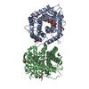
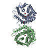
| Deposited unit | 
| ||||||||
|---|---|---|---|---|---|---|---|---|---|
| 1 | 
| ||||||||
| Unit cell |
| ||||||||
| Details | Dimer is generated by: -x, y, -z+1/2 |
-Components
| #1: Protein | Mass: 37601.570 Da / Num. of mol.: 1 / Fragment: Catalytic domain (residues 103-427) Source method: isolated from a genetically manipulated source Source: (gene. exp.) Homo sapiens (human) / Gene: PAH / Plasmid: pMAL / Production host:  |
|---|---|
| #2: Chemical | ChemComp-FE2 /   Mass: 55.845 Da / Num. of mol.: 1 / Source method: obtained synthetically / Formula: Fe Mass: 55.845 Da / Num. of mol.: 1 / Source method: obtained synthetically / Formula: Fe |
| #3: Chemical | ChemComp-H4B /   Mass: 241.247 Da / Num. of mol.: 1 / Source method: obtained synthetically / Formula: C9H15N5O3 / Comment: neurotransmitter*YM Mass: 241.247 Da / Num. of mol.: 1 / Source method: obtained synthetically / Formula: C9H15N5O3 / Comment: neurotransmitter*YM |
| #4: Chemical | ChemComp-TIH /   Type: L-peptide linking / Mass: 171.217 Da / Num. of mol.: 1 / Source method: obtained synthetically / Formula: C7H9NO2S Type: L-peptide linking / Mass: 171.217 Da / Num. of mol.: 1 / Source method: obtained synthetically / Formula: C7H9NO2S |
| #5: Water | ChemComp-HOH /  Mass: 18.015 Da / Num. of mol.: 39 / Source method: isolated from a natural source / Formula: H2O Mass: 18.015 Da / Num. of mol.: 39 / Source method: isolated from a natural source / Formula: H2O |
-Experimental details
-Experiment
| Experiment | Method: X-RAY DIFFRACTION / Number of used crystals: 1 |
|---|
- Sample preparation
| Crystal | Density Matthews: 2.85 Å3/Da / Density % sol: 56.89 % | ||||||||||||||||||
|---|---|---|---|---|---|---|---|---|---|---|---|---|---|---|---|---|---|---|---|
| Crystal grow | Temperature: 277 K / Method: vapor diffusion, hanging drop / pH: 6.8 Details: 20% PEG 2000, 10% ethylene glycol 0.12M Na-Hepes pH 6.8, 30mM Na-dithionite, 10mM BH4, VAPOR DIFFUSION, HANGING DROP, temperature 277K | ||||||||||||||||||
| Crystal grow | *PLUS Method: vapor diffusion | ||||||||||||||||||
| Components of the solutions | *PLUS
|
-Data collection
| Diffraction | Mean temperature: 100 K |
|---|---|
| Diffraction source | Source: SYNCHROTRON / Site: ESRF  / Beamline: BM1A / Wavelength: 0.8 Å / Beamline: BM1A / Wavelength: 0.8 Å |
| Detector | Type: MAR scanner 345 mm plate / Detector: IMAGE PLATE / Date: Apr 8, 2001 / Details: mirrors |
| Radiation | Protocol: SINGLE WAVELENGTH / Monochromatic (M) / Laue (L): M / Scattering type: x-ray |
| Radiation wavelength | Wavelength: 0.8 Å / Relative weight: 1 |
| Reflection | Resolution: 2.5→10 Å / Num. all: 14178 / Num. obs: 14178 / % possible obs: 94.4 % / Observed criterion σ(F): 0 / Observed criterion σ(I): 0 / Redundancy: 4.2 % / Biso Wilson estimate: 41.3 Å2 / Rmerge(I) obs: 0.092 / Rsym value: 0.092 / Net I/σ(I): 7.1 |
| Reflection shell | Resolution: 2.5→2.64 Å / Redundancy: 4.1 % / Rmerge(I) obs: 0.377 / Mean I/σ(I) obs: 2.1 / Num. unique all: 2071 / Rsym value: 0.377 / % possible all: 95.2 |
| Reflection | *PLUS Highest resolution: 2.5 Å / Lowest resolution: 10 Å / Num. measured all: 59237 |
| Reflection shell | *PLUS % possible obs: 95.2 % / Num. unique obs: 2071 / Num. measured obs: 8578 |
- Processing
| Software |
| |||||||||||||||||||||||||
|---|---|---|---|---|---|---|---|---|---|---|---|---|---|---|---|---|---|---|---|---|---|---|---|---|---|---|
| Refinement | Method to determine structure: adopting entry 1PAH phases Starting model: PDB entry 1PAH Resolution: 2.5→10 Å / Isotropic thermal model: isotropic / Cross valid method: THROUGHOUT / σ(F): 0 / σ(I): 0 / Stereochemistry target values: Engh & Huber
| |||||||||||||||||||||||||
| Displacement parameters | Biso mean: 34.3 Å2 | |||||||||||||||||||||||||
| Refine analyze | Luzzati coordinate error obs: 0.35 Å / Luzzati sigma a obs: 0.29 Å | |||||||||||||||||||||||||
| Refinement step | Cycle: LAST / Resolution: 2.5→10 Å
| |||||||||||||||||||||||||
| Refine LS restraints |
| |||||||||||||||||||||||||
| LS refinement shell | Resolution: 2.5→2.53 Å /
| |||||||||||||||||||||||||
| Refinement | *PLUS Lowest resolution: 10 Å / % reflection Rfree: 10 % / Rfactor Rwork: 0.22 | |||||||||||||||||||||||||
| Solvent computation | *PLUS | |||||||||||||||||||||||||
| Displacement parameters | *PLUS | |||||||||||||||||||||||||
| Refine LS restraints | *PLUS
|
