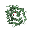
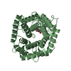
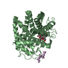
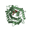
[English] 日本語
Yorodumi
- PDB-5m77: a GH76 family enzyme structure -

+
Open data


ID or keywords:

Loading...

-
Basic information

Entry
Database: PDB / ID: 5m77
Titlea GH76 family enzyme structure
ComponentsAlpha-1,6-mannanase
KeywordsHYDROLASE / glycoside HYDROLASE / complex / mannanase / S-linked polysaccharide
Function / homology
Function and homology information


catalytic activity / carbohydrate binding / carbohydrate metabolic process
Similarity search - Function
Glycoside hydrolase, family 76 / Glycosyl hydrolase family 76 / Cellulose binding, type IV / Cellulose Binding Domain Type IV / Carbohydrate binding module (family 6) / CBM6 (carbohydrate binding type-6) domain profile. / Carbohydrate binding module family 6 / Glycosyltransferase - #20 / Six-hairpin glycosidase superfamily / Glycosyltransferase ...Glycoside hydrolase, family 76 / Glycosyl hydrolase family 76 / Cellulose binding, type IV / Cellulose Binding Domain Type IV / Carbohydrate binding module (family 6) / CBM6 (carbohydrate binding type-6) domain profile. / Carbohydrate binding module family 6 / Glycosyltransferase - #20 / Six-hairpin glycosidase superfamily / Glycosyltransferase / Alpha/alpha barrel / Galactose-binding-like domain superfamily / Mainly Alpha
Similarity search - Domain/homology
Biological speciesBacillus circulans (bacteria)
MethodX-RAY DIFFRACTION / SYNCHROTRON / MOLECULAR REPLACEMENT / Resolution: 1.46 Å
AuthorsJin, Y. / Williams, S. / Davies, G.
Funding support United Kingdom, 1items
OrganizationGrant numberCountry
European Research CouncilAdG-322942 United Kingdom
CitationJournal: Chem. Commun. (Camb.) / Year: 2017
Title: An atypical interaction explains the high-affinity of a non-hydrolyzable S-linked 1,6-alpha-mannanase inhibitor.
Authors: Belz, T. / Jin, Y. / Coines, J. / Rovira, C. / Davies, G.J. / Williams, S.J.
History
DepositionOct 26, 2016Deposition site: PDBE / Processing site: PDBE
Revision 1.0Aug 9, 2017Provider: repository / Type: Initial release
Revision 1.1Aug 16, 2017Group: Database references / Category: citation / citation_author
Item: _citation.journal_abbrev / _citation.pdbx_database_id_DOI ..._citation.journal_abbrev / _citation.pdbx_database_id_DOI / _citation.pdbx_database_id_PubMed / _citation.title
Revision 1.2Aug 23, 2017Group: Database references / Category: citation
Item: _citation.journal_volume / _citation.page_first / _citation.page_last
Revision 2.0Jul 29, 2020Group: Atomic model / Author supporting evidence ...Atomic model / Author supporting evidence / Data collection / Derived calculations / Structure summary
Category: atom_site / atom_site_anisotrop ...atom_site / atom_site_anisotrop / chem_comp / entity / pdbx_audit_support / pdbx_branch_scheme / pdbx_entity_branch / pdbx_entity_branch_descriptor / pdbx_entity_branch_link / pdbx_entity_branch_list / pdbx_entity_nonpoly / pdbx_nonpoly_scheme / pdbx_struct_assembly_gen / struct_asym / struct_conn / struct_site / struct_site_gen
Item: _atom_site.B_iso_or_equiv / _atom_site.Cartn_x ..._atom_site.B_iso_or_equiv / _atom_site.Cartn_x / _atom_site.Cartn_y / _atom_site.Cartn_z / _atom_site.auth_asym_id / _atom_site.auth_atom_id / _atom_site.auth_comp_id / _atom_site.auth_seq_id / _atom_site.label_asym_id / _atom_site.label_atom_id / _atom_site.label_comp_id / _atom_site.label_entity_id / _atom_site.occupancy / _atom_site.type_symbol / _atom_site_anisotrop.U[1][1] / _atom_site_anisotrop.U[1][2] / _atom_site_anisotrop.U[1][3] / _atom_site_anisotrop.U[2][2] / _atom_site_anisotrop.U[2][3] / _atom_site_anisotrop.U[3][3] / _atom_site_anisotrop.pdbx_auth_asym_id / _atom_site_anisotrop.pdbx_auth_atom_id / _atom_site_anisotrop.pdbx_auth_comp_id / _atom_site_anisotrop.pdbx_auth_seq_id / _atom_site_anisotrop.pdbx_label_asym_id / _atom_site_anisotrop.pdbx_label_atom_id / _atom_site_anisotrop.pdbx_label_comp_id / _atom_site_anisotrop.type_symbol / _chem_comp.name / _chem_comp.type / _pdbx_audit_support.funding_organization / _pdbx_struct_assembly_gen.asym_id_list / _struct_conn.ptnr1_auth_asym_id / _struct_conn.ptnr1_auth_seq_id / _struct_conn.ptnr1_label_asym_id / _struct_conn.ptnr1_label_atom_id / _struct_conn.ptnr2_auth_asym_id / _struct_conn.ptnr2_auth_seq_id / _struct_conn.ptnr2_label_asym_id
Description: Carbohydrate remediation / Provider: repository / Type: Remediation
Revision 2.1Jan 17, 2024Group: Data collection / Database references ...Data collection / Database references / Refinement description / Structure summary
Category: chem_comp / chem_comp_atom ...chem_comp / chem_comp_atom / chem_comp_bond / database_2 / pdbx_initial_refinement_model / struct_ncs_dom_lim
Item: _chem_comp.pdbx_synonyms / _database_2.pdbx_DOI ..._chem_comp.pdbx_synonyms / _database_2.pdbx_DOI / _database_2.pdbx_database_accession / _struct_ncs_dom_lim.beg_auth_comp_id / _struct_ncs_dom_lim.beg_label_asym_id / _struct_ncs_dom_lim.beg_label_comp_id / _struct_ncs_dom_lim.beg_label_seq_id / _struct_ncs_dom_lim.end_auth_comp_id / _struct_ncs_dom_lim.end_label_asym_id / _struct_ncs_dom_lim.end_label_comp_id / _struct_ncs_dom_lim.end_label_seq_id

-
Structure visualization

Structure viewerMolecule:
MolmilJmol/JSmol

Downloads & links

-
Assembly

Deposited unit
A: Alpha-1,6-mannanase
B: Alpha-1,6-mannanase
hetero molecules


Theoretical massNumber of molelcules
Total (without water)133,24119
Polymers131,3032
Non-polymers1,93817
Water8,107450
1
A: Alpha-1,6-mannanase
hetero molecules


Theoretical massNumber of molelcules
Total (without water)66,5278
Polymers65,6511
Non-polymers8767
Water181
TypeNameSymmetry operationNumber
identity operation1_555x,y,z1
MethodPISA
2
B: Alpha-1,6-mannanase
hetero molecules


Theoretical massNumber of molelcules
Total (without water)66,71411
Polymers65,6511
Non-polymers1,06210
Water181
TypeNameSymmetry operationNumber
identity operation1_555x,y,z1
MethodPISA
Unit cell
Length a, b, c (Å)84.060, 85.170, 101.860
Angle α, β, γ (deg.)90.00, 90.00, 90.00
Int Tables number19
Space group name H-MP212121
Noncrystallographic symmetry (NCS)NCS domain:
IDEns-IDDetails
11A
21B

NCS domain segments:

Component-ID: _ / Ens-ID: 1 / Beg auth comp-ID: SER / Beg label comp-ID: SER / End auth comp-ID: ILE / End label comp-ID: ILE / Refine code: _ / Auth seq-ID: 39 - 375 / Label seq-ID: 39 - 375

Dom-IDAuth asym-IDLabel asym-ID
1AA
2BB

-
Components

#1: Protein Alpha-1,6-mannanase


Mass: 65651.320 Da / Num. of mol.: 2 / Mutation: R341Q
Source method: isolated from a genetically manipulated source
Source: (gene. exp.) Bacillus circulans (bacteria) / Gene: aman6 / Production host: Escherichia coli (E. coli) / References: UniProt: Q9Z4P9
#2: Polysaccharide 1-thio-alpha-D-mannopyranose-(1-6)-[(3S,4R,5R)-4,5-dihydroxypiperidin-3-yl]methyl 1-thio-alpha-D- ...1-thio-alpha-D-mannopyranose-(1-6)-[(3S,4R,5R)-4,5-dihydroxypiperidin-3-yl]methyl 1-thio-alpha-D-mannopyranoside


Type: oligosaccharide / Mass: 503.585 Da / Num. of mol.: 2 / Source method: obtained synthetically
DescriptorTypeProgram
[][<C6N1O2>]{[(1+S)][a-D-Manp1SH6SH]{[(6+S)][a-D-Manp]{}}}LINUCSPDB-CARE
#3: Chemical
ChemComp-EDO / 1,2-ETHANEDIOL / ETHYLENE GLYCOL


Mass: 62.068 Da / Num. of mol.: 15 / Source method: obtained synthetically / Formula: C2H6O2
#4: Water ChemComp-HOH / water


Mass: 18.015 Da / Num. of mol.: 450 / Source method: isolated from a natural source / Formula: H2O

-
Experimental details

-
Experiment

ExperimentMethod: X-RAY DIFFRACTION / Number of used crystals: 1

-
Sample preparation

Crystal growTemperature: 293 K / Method: vapor diffusion / pH: 6.5 / Details: 0.1 M AMMONIUM NITRATE, PH 6.5, 20% PEG 3350

-
Data collection

DiffractionMean temperature: 100 K
Diffraction sourceSource: SYNCHROTRON / Site: Diamond / Beamline: I02 / Wavelength: 0.97959 Å
DetectorType: DECTRIS PILATUS 6M-F / Detector: PIXEL / Date: Mar 6, 2016
RadiationProtocol: SINGLE WAVELENGTH / Monochromatic (M) / Laue (L): M / Scattering type: x-ray
Radiation wavelengthWavelength: 0.97959 Å / Relative weight: 1
ReflectionResolution: 1.46→50.93 Å / Num. obs: 127095 / % possible obs: 99.9 % / Observed criterion σ(I): 1.4 / Redundancy: 6.5 % / CC1/2: 0.999 / Rmerge(I) obs: 0.05 / Net I/σ(I): 16.2
Reflection shellResolution: 1.46→1.5 Å / Redundancy: 5.8 % / Mean I/σ(I) obs: 1.4 / Num. unique all: 9293 / CC1/2: 0.531 / % possible all: 99.6

-
Processing

Software
NameVersionClassification
REFMAC5.8.0155refinement
xia2data reduction
xia2data scaling
MOLREPphasing
RefinementMethod to determine structure: MOLECULAR REPLACEMENT
Starting model: 4d4a
Resolution: 1.46→50.93 Å / Cor.coef. Fo:Fc: 0.983 / Cor.coef. Fo:Fc free: 0.974 / SU B: 2.504 / SU ML: 0.04 / Cross valid method: THROUGHOUT / ESU R: 0.053 / ESU R Free: 0.055 / Details: HYDROGENS HAVE BEEN ADDED IN THE RIDING POSITIONS
RfactorNum. reflection% reflectionSelection details
Rfree0.16524 6317 5 %RANDOM
Rwork0.12067 ---
obs0.12288 127007 99.86 %-
Solvent computationIon probe radii: 0.8 Å / Shrinkage radii: 0.8 Å / VDW probe radii: 1.2 Å
Displacement parametersBiso mean: 26.791 Å2
Baniso -1Baniso -2Baniso -3
1-0.5 Å2-0 Å20 Å2
2---0.49 Å2-0 Å2
3----0.01 Å2
Refinement stepCycle: 1 / Resolution: 1.46→50.93 Å
ProteinNucleic acidLigandSolventTotal
Num. atoms5341 0 124 450 5915
Refine LS restraints
Refine-IDTypeDev idealDev ideal targetNumber
X-RAY DIFFRACTIONr_bond_refined_d0.0190.0195644
X-RAY DIFFRACTIONr_bond_other_d0.0020.024986
X-RAY DIFFRACTIONr_angle_refined_deg1.7411.9137659
X-RAY DIFFRACTIONr_angle_other_deg1.08311460
X-RAY DIFFRACTIONr_dihedral_angle_1_deg5.3955674
X-RAY DIFFRACTIONr_dihedral_angle_2_deg37.23325.333300
X-RAY DIFFRACTIONr_dihedral_angle_3_deg11.49315840
X-RAY DIFFRACTIONr_dihedral_angle_4_deg24.6831513
X-RAY DIFFRACTIONr_chiral_restr0.120.2789
X-RAY DIFFRACTIONr_gen_planes_refined0.010.026505
X-RAY DIFFRACTIONr_gen_planes_other0.0030.021404
X-RAY DIFFRACTIONr_nbd_refined
X-RAY DIFFRACTIONr_nbd_other
X-RAY DIFFRACTIONr_nbtor_refined
X-RAY DIFFRACTIONr_nbtor_other
X-RAY DIFFRACTIONr_xyhbond_nbd_refined
X-RAY DIFFRACTIONr_xyhbond_nbd_other
X-RAY DIFFRACTIONr_metal_ion_refined
X-RAY DIFFRACTIONr_metal_ion_other
X-RAY DIFFRACTIONr_symmetry_vdw_refined
X-RAY DIFFRACTIONr_symmetry_vdw_other
X-RAY DIFFRACTIONr_symmetry_hbond_refined
X-RAY DIFFRACTIONr_symmetry_hbond_other
X-RAY DIFFRACTIONr_symmetry_metal_ion_refined
X-RAY DIFFRACTIONr_symmetry_metal_ion_other
X-RAY DIFFRACTIONr_mcbond_it3.1532.412681
X-RAY DIFFRACTIONr_mcbond_other3.1532.4092680
X-RAY DIFFRACTIONr_mcangle_it3.3083.6273348
X-RAY DIFFRACTIONr_mcangle_other3.3083.6273349
X-RAY DIFFRACTIONr_scbond_it4.4842.7422963
X-RAY DIFFRACTIONr_scbond_other4.4842.7422963
X-RAY DIFFRACTIONr_scangle_it
X-RAY DIFFRACTIONr_scangle_other4.8333.9784308
X-RAY DIFFRACTIONr_long_range_B_refined4.53229.676864
X-RAY DIFFRACTIONr_long_range_B_other4.53229.676864
X-RAY DIFFRACTIONr_rigid_bond_restr3.992310630
X-RAY DIFFRACTIONr_sphericity_free26.0075297
X-RAY DIFFRACTIONr_sphericity_bonded15.387510638
Refine LS restraints NCS

Ens-ID: 1 / Number: 23622 / Refine-ID: X-RAY DIFFRACTION / Type: interatomic distance / Rms dev position: 0.07 Å / Weight position: 0.05

Dom-IDAuth asym-ID
1A
2B
LS refinement shellResolution: 1.46→1.498 Å / Total num. of bins used: 20
RfactorNum. reflection% reflection
Rfree0.29 471 -
Rwork0.254 8812 -
obs-6317 99.44 %

+
About Yorodumi

-
News

-
Feb 9, 2022. New format data for meta-information of EMDB entries

New format data for meta-information of EMDB entries

  • Version 3 of the EMDB header file is now the official format.
  • The previous official version 1.9 will be removed from the archive.

Related info.:EMDB header

External links:wwPDB to switch to version 3 of the EMDB data model

-
Aug 12, 2020. Covid-19 info

Covid-19 info

URL: https://pdbjlvh1.pdbj.org/emnavi/covid19.php

New page: Covid-19 featured information page in EM Navigator.

Related info.:Covid-19 info / Mar 5, 2020. Novel coronavirus structure data

+
Mar 5, 2020. Novel coronavirus structure data

Novel coronavirus structure data

Related info.:Yorodumi Speices / Aug 12, 2020. Covid-19 info

External links:COVID-19 featured content - PDBj / Molecule of the Month (242):Coronavirus Proteases

+
Jan 31, 2019. EMDB accession codes are about to change! (news from PDBe EMDB page)

EMDB accession codes are about to change! (news from PDBe EMDB page)

  • The allocation of 4 digits for EMDB accession codes will soon come to an end. Whilst these codes will remain in use, new EMDB accession codes will include an additional digit and will expand incrementally as the available range of codes is exhausted. The current 4-digit format prefixed with “EMD-” (i.e. EMD-XXXX) will advance to a 5-digit format (i.e. EMD-XXXXX), and so on. It is currently estimated that the 4-digit codes will be depleted around Spring 2019, at which point the 5-digit format will come into force.
  • The EM Navigator/Yorodumi systems omit the EMD- prefix.

Related info.:Q: What is EMD? / ID/Accession-code notation in Yorodumi/EM Navigator

External links:EMDB Accession Codes are Changing Soon! / Contact to PDBj

+
Jul 12, 2017. Major update of PDB

Major update of PDB

  • wwPDB released updated PDB data conforming to the new PDBx/mmCIF dictionary.
  • This is a major update changing the version number from 4 to 5, and with Remediation, in which all the entries are updated.
  • In this update, many items about electron microscopy experimental information are reorganized (e.g. em_software).
  • Now, EM Navigator and Yorodumi are based on the updated data.

External links:wwPDB Remediation / Enriched Model Files Conforming to OneDep Data Standards Now Available in the PDB FTP Archive

-
Yorodumi

Thousand views of thousand structures

  • Yorodumi is a browser for structure data from EMDB, PDB, SASBDB, etc.
  • This page is also the successor to EM Navigator detail page, and also detail information page/front-end page for Omokage search.
  • The word "yorodu" (or yorozu) is an old Japanese word meaning "ten thousand". "mi" (miru) is to see.

Related info.:EMDB / PDB / SASBDB / Comparison of 3 databanks / Yorodumi Search / Aug 31, 2016. New EM Navigator & Yorodumi / Yorodumi Papers / Jmol/JSmol / Function and homology information / Changes in new EM Navigator and Yorodumi

Read more