 Movie
Movie Controller

[English] 日本語
 Yorodumi
- PDB-1kqd: Structure of Nitroreductase from E. cloacae Bound with 2e-Reduced... -
+ Open data

- Basic information
| Entry | Database: PDB / ID: 1kqd | ||||||
|---|---|---|---|---|---|---|---|
| Title | Structure of Nitroreductase from E. cloacae Bound with 2e-Reduced Flavin Mononucleotide (FMN) | ||||||
 Components Components | OXYGEN-INSENSITIVE NAD(P)H NITROREDUCTASE | ||||||
 Keywords Keywords | OXIDOREDUCTASE / nitroreductase / reduced hydroquinone / flavin | ||||||
| Function / homology |  Function and homology information Function and homology informationoxidoreductase activity, acting on other nitrogenous compounds as donors, with NAD or NADP as acceptor / 2,4,6-trinitrotoluene catabolic process / Oxidoreductases / cytosol Similarity search - Function | ||||||
| Biological species |  Enterobacter cloacae (bacteria) Enterobacter cloacae (bacteria) | ||||||
| Method |  X-RAY DIFFRACTION / MOLECULAR REPLACEMENT / Resolution: 1.9 Å X-RAY DIFFRACTION / MOLECULAR REPLACEMENT / Resolution: 1.9 Å | ||||||
 Authors Authors | Haynes, C.A. / Koder, R.L. / Miller, A.F. / Rodgers, D.W. | ||||||
 Citation Citation | Journal: J.Biol.Chem. / Year: 2002 Title: Structures of nitroreductase in three states: effects of inhibitor binding and reduction. Authors: Haynes, C.A. / Koder, R.L. / Miller, A.F. / Rodgers, D.W. | ||||||
| History |
|
- Structure visualization
| Structure viewer | Molecule: MolmilJmol/JSmol |
|---|
- Downloads & links
-Download
| PDBx/mmCIF format | 1kqd.cif.gz | 185.1 KB | Display | PDBx/mmCIF format |
|---|---|---|---|---|
| PDB format | pdb1kqd.ent.gz | 148.4 KB | Display | PDB format |
| PDBx/mmJSON format | 1kqd.json.gz | Tree view | PDBx/mmJSON format | |
| Others |  Other downloads Other downloads |
-Validation report
| Arichive directory | https://data.pdbj.org/pub/pdb/validation_reports/kq/1kqdftp://data.pdbj.org/pub/pdb/validation_reports/kq/1kqd | HTTPS FTP |
|---|
-Related structure data
-Links
 PDBj
PDBj- Assembly
| Deposited unit | 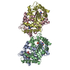
| ||||||||
|---|---|---|---|---|---|---|---|---|---|
| 1 | 
| ||||||||
| 2 | 
| ||||||||
| Unit cell |
| ||||||||
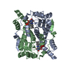
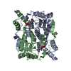
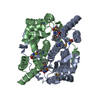
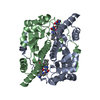
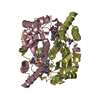
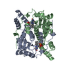
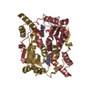
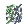
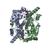
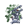
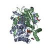
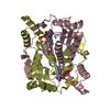
| Details | Dimer. Chains A and B represent one dimer, chains C and D represent the other dimer; both dimers are in the asymmetric unit. We are depositing all four monomers. |
-Components
| #1: Protein | Mass: 23982.178 Da / Num. of mol.: 4 Source method: isolated from a genetically manipulated source Source: (gene. exp.) Enterobacter cloacae (bacteria) / Plasmid: pET24d(+) / Species (production host): Escherichia coli / Production host: #2: Chemical | ChemComp-FMN /   Mass: 456.344 Da / Num. of mol.: 4 / Source method: obtained synthetically / Formula: C17H21N4O9P Mass: 456.344 Da / Num. of mol.: 4 / Source method: obtained synthetically / Formula: C17H21N4O9P#3: Water | ChemComp-HOH / |  Mass: 18.015 Da / Num. of mol.: 437 / Source method: isolated from a natural source / Formula: H2O Mass: 18.015 Da / Num. of mol.: 437 / Source method: isolated from a natural source / Formula: H2O |
|---|
-Experimental details
-Experiment
| Experiment | Method: X-RAY DIFFRACTION / Number of used crystals: 1 |
|---|
- Sample preparation
| Crystal | Density Matthews: 2.14 Å3/Da / Density % sol: 42.44 % | |||||||||||||||||||||||||||||||||||||||||||||||||
|---|---|---|---|---|---|---|---|---|---|---|---|---|---|---|---|---|---|---|---|---|---|---|---|---|---|---|---|---|---|---|---|---|---|---|---|---|---|---|---|---|---|---|---|---|---|---|---|---|---|---|
| Crystal grow | Temperature: 277 K / Method: vapor diffusion, hanging drop / pH: 4.8 Details: homopipes, acetate, PEG 4000, pH 4.8, VAPOR DIFFUSION, HANGING DROP, temperature 277K | |||||||||||||||||||||||||||||||||||||||||||||||||
| Crystal grow | *PLUS Temperature: 4 ℃ / pH: 7 | |||||||||||||||||||||||||||||||||||||||||||||||||
| Components of the solutions | *PLUS
|
-Data collection
| Diffraction | Mean temperature: 115 K |
|---|---|
| Diffraction source | Source: ROTATING ANODE / Type: RIGAKU RU200 / Wavelength: 1.5418 |
| Detector | Type: RIGAKU RAXIS IV / Detector: IMAGE PLATE / Date: Nov 25, 2000 / Details: graded multi-layer |
| Radiation | Monochromator: Ni Filter / Protocol: SINGLE WAVELENGTH / Monochromatic (M) / Laue (L): M / Scattering type: x-ray |
| Radiation wavelength | Wavelength: 1.5418 Å / Relative weight: 1 |
| Reflection | Resolution: 1.9→20 Å / Num. all: 63677 / Num. obs: 62505 / % possible obs: 0.982 % / Observed criterion σ(F): 0 / Observed criterion σ(I): -3 / Redundancy: 4 % / Rmerge(I) obs: 0.054 / Net I/σ(I): 27.6 |
| Reflection shell | Resolution: 1.9→1.99 Å / Redundancy: 3.09 % / Rmerge(I) obs: 0.143 / Mean I/σ(I) obs: 7.5 / % possible all: 0.889 |
| Reflection | *PLUS Lowest resolution: 20 Å / % possible obs: 97.7 % / Redundancy: 4 % |
| Reflection shell | *PLUS % possible obs: 88.9 % |
- Processing
| Software |
| |||||||||||||||||||||||||
|---|---|---|---|---|---|---|---|---|---|---|---|---|---|---|---|---|---|---|---|---|---|---|---|---|---|---|
| Refinement | Method to determine structure: MOLECULAR REPLACEMENT / Resolution: 1.9→20 Å / σ(F): 0 / σ(I): 0 / Stereochemistry target values: Engh & Huber
| |||||||||||||||||||||||||
| Refinement step | Cycle: LAST / Resolution: 1.9→20 Å
| |||||||||||||||||||||||||
| Refine LS restraints |
| |||||||||||||||||||||||||
| Software | *PLUS Name: CNS / Classification: refinement | |||||||||||||||||||||||||
| Refinement | *PLUS σ(F): 0 / % reflection Rfree: 10 % / Rfactor obs: 0.188 / Rfactor Rfree: 0.22 | |||||||||||||||||||||||||
| Solvent computation | *PLUS | |||||||||||||||||||||||||
| Displacement parameters | *PLUS | |||||||||||||||||||||||||
| Refine LS restraints | *PLUS
|
