 Movie
Movie Controller

[English] 日本語
 Yorodumi
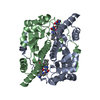
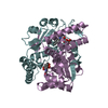
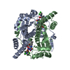
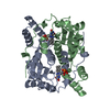
- PDB-1ylu: The structure of E. coli nitroreductase with bound acetate, cryst... -
+ Open data

- Basic information
| Entry | Database: PDB / ID: 1ylu | ||||||
|---|---|---|---|---|---|---|---|
| Title | The structure of E. coli nitroreductase with bound acetate, crystal form 2 | ||||||
 Components Components | Oxygen-insensitive NAD(P)H nitroreductase | ||||||
 Keywords Keywords | OXIDOREDUCTASE / Oxygen-insensitive NAD(P)H nitroreductase | ||||||
| Function / homology |  Function and homology information Function and homology information6,7-dihydropteridine reductase / 6,7-dihydropteridine reductase activity / 2,4,6-trinitrotoluene catabolic process / NAD(P)H dehydrogenase (quinone) activity / Oxidoreductases / FMN binding / oxidoreductase activity / protein homodimerization activity / membrane / identical protein binding / cytosol Similarity search - Function | ||||||
| Biological species |  | ||||||
| Method |  X-RAY DIFFRACTION / SYNCHROTRON / MOLECULAR REPLACEMENT / Resolution: 2 Å X-RAY DIFFRACTION / SYNCHROTRON / MOLECULAR REPLACEMENT / Resolution: 2 Å | ||||||
 Authors Authors | Race, P.R. / Lovering, A.L. / Green, R.M. / Ossor, A. / White, S.A. / Searle, P.F. / Wrighton, C.J. / Hyde, E.I. | ||||||
 Citation Citation | Journal: J.Biol.Chem. / Year: 2005 Title: Structural and mechanistic studies of Escherichia coli nitroreductase with the antibiotic nitrofurazone. Reversed binding orientations in different redox states of the enzyme. Authors: Race, P.R. / Lovering, A.L. / Green, R.M. / Ossor, A. / White, S.A. / Searle, P.F. / Wrighton, C.J. / Hyde, E.I. #1: Journal: J.Mol.Biol. / Year: 2001Title: The Structure of Escherichia coli Nitroreductase Complexed with Nicotinic Acid: Three Crystal Forms at 1.7 A, 1.8 A and 2.4 A Resolution Authors: Lovering, A.L. / Hyde, E.I. / Searle, P.F. / White, S.A. | ||||||
| History |
| ||||||
| Remark 600 | HETEROGEN LIGANDS 1218-1219 ARE ACCOSIATED WITH CHAIN A. LIGANDS 2218-2219 ARE ACCOSIATED WITH CHAIN B. |
- Structure visualization
| Structure viewer | Molecule: MolmilJmol/JSmol |
|---|
- Downloads & links
-Download
| PDBx/mmCIF format | 1ylu.cif.gz | 111.7 KB | Display | PDBx/mmCIF format |
|---|---|---|---|---|
| PDB format | pdb1ylu.ent.gz | 84.5 KB | Display | PDB format |
| PDBx/mmJSON format | 1ylu.json.gz | Tree view | PDBx/mmJSON format | |
| Others |  Other downloads Other downloads |
-Validation report
| Arichive directory | https://data.pdbj.org/pub/pdb/validation_reports/yl/1yluftp://data.pdbj.org/pub/pdb/validation_reports/yl/1ylu | HTTPS FTP |
|---|
-Related structure data
| Related structure data |  1ykiC  1ylrC  1icvS S: Starting model for refinement C: citing same article ( |
|---|---|
| Similar structure data |


-Links
 PDBj
PDBj
- Assembly
| Deposited unit | 
| |||||||||
|---|---|---|---|---|---|---|---|---|---|---|
| 1 |
| |||||||||
| Unit cell |
| |||||||||
| Components on special symmetry positions |
|
-Components
| #1: Protein | Mass: 23937.182 Da / Num. of mol.: 2 Source method: isolated from a genetically manipulated source Source: (gene. exp.) #2: Chemical |   Mass: 59.044 Da / Num. of mol.: 3 / Source method: obtained synthetically / Formula: C2H3O2 Mass: 59.044 Da / Num. of mol.: 3 / Source method: obtained synthetically / Formula: C2H3O2#3: Chemical |   Mass: 456.344 Da / Num. of mol.: 2 / Source method: obtained synthetically / Formula: C17H21N4O9P Mass: 456.344 Da / Num. of mol.: 2 / Source method: obtained synthetically / Formula: C17H21N4O9P#4: Water | ChemComp-HOH / |  Mass: 18.015 Da / Num. of mol.: 528 / Source method: isolated from a natural source / Formula: H2O Mass: 18.015 Da / Num. of mol.: 528 / Source method: isolated from a natural source / Formula: H2O |
|---|
-Experimental details
-Experiment
| Experiment | Method: X-RAY DIFFRACTION / Number of used crystals: 1 |
|---|
- Sample preparation
| Crystal | Density Matthews: 2 Å3/Da / Density % sol: 36.8 % |
|---|---|
| Crystal grow | Temperature: 291 K / Method: vapor diffusion, hanging drop / pH: 4.6 Details: PEG4000, ethylene glycol, nicotinic acid, sodium acetate, pH 4.6, VAPOR DIFFUSION, HANGING DROP, temperature 291K |
-Data collection
| Diffraction | Mean temperature: 100 K |
|---|---|
| Diffraction source | Source: SYNCHROTRON / Site: ESRF  / Beamline: BM14 / Wavelength: 0.97626 Å / Beamline: BM14 / Wavelength: 0.97626 Å |
| Detector | Type: ADSC QUANTUM 4 / Detector: CCD / Date: Nov 11, 2000 |
| Radiation | Protocol: SINGLE WAVELENGTH / Monochromatic (M) / Laue (L): M / Scattering type: x-ray |
| Radiation wavelength | Wavelength: 0.97626 Å / Relative weight: 1 |
| Reflection | Resolution: 2→26.93 Å / Num. all: 26207 / Num. obs: 26207 / % possible obs: 95.6 % / Observed criterion σ(F): 1 / Observed criterion σ(I): 1 / Redundancy: 3 % / Biso Wilson estimate: 15.9 Å2 / Rsym value: 0.071 / Net I/σ(I): 13.1 |
| Reflection shell | Resolution: 2→2.11 Å / Redundancy: 2.6 % / Mean I/σ(I) obs: 5.4 / Num. unique all: 3743 / Rsym value: 0.173 / % possible all: 95.7 |
- Processing
| Software |
| ||||||||||||||||||||||||||||||||||||||||||||||||||||||||||||||||||||||||||||||||||||||||||||||||||||||||||||||||||||||||||||||||||||||||||||||||||||||||||||||||||||||||||
|---|---|---|---|---|---|---|---|---|---|---|---|---|---|---|---|---|---|---|---|---|---|---|---|---|---|---|---|---|---|---|---|---|---|---|---|---|---|---|---|---|---|---|---|---|---|---|---|---|---|---|---|---|---|---|---|---|---|---|---|---|---|---|---|---|---|---|---|---|---|---|---|---|---|---|---|---|---|---|---|---|---|---|---|---|---|---|---|---|---|---|---|---|---|---|---|---|---|---|---|---|---|---|---|---|---|---|---|---|---|---|---|---|---|---|---|---|---|---|---|---|---|---|---|---|---|---|---|---|---|---|---|---|---|---|---|---|---|---|---|---|---|---|---|---|---|---|---|---|---|---|---|---|---|---|---|---|---|---|---|---|---|---|---|---|---|---|---|---|---|---|---|
| Refinement | Method to determine structure: MOLECULAR REPLACEMENT Starting model: pdb entry 1ICV Resolution: 2→53.45 Å / Cor.coef. Fo:Fc: 0.962 / Cor.coef. Fo:Fc free: 0.928 / SU B: 3.634 / SU ML: 0.102 / Isotropic thermal model: isotropic / Cross valid method: THROUGHOUT / σ(F): 0 / ESU R: 0.196 / ESU R Free: 0.167 / Stereochemistry target values: Engh & Huber / Details: HYDROGENS HAVE BEEN ADDED IN THE RIDING POSITIONS
| ||||||||||||||||||||||||||||||||||||||||||||||||||||||||||||||||||||||||||||||||||||||||||||||||||||||||||||||||||||||||||||||||||||||||||||||||||||||||||||||||||||||||||
| Solvent computation | Ion probe radii: 0.8 Å / Shrinkage radii: 0.8 Å / VDW probe radii: 1.4 Å / Solvent model: BABINET MODEL WITH MASK | ||||||||||||||||||||||||||||||||||||||||||||||||||||||||||||||||||||||||||||||||||||||||||||||||||||||||||||||||||||||||||||||||||||||||||||||||||||||||||||||||||||||||||
| Displacement parameters | Biso mean: 10.381 Å2
| ||||||||||||||||||||||||||||||||||||||||||||||||||||||||||||||||||||||||||||||||||||||||||||||||||||||||||||||||||||||||||||||||||||||||||||||||||||||||||||||||||||||||||
| Refinement step | Cycle: LAST / Resolution: 2→53.45 Å
| ||||||||||||||||||||||||||||||||||||||||||||||||||||||||||||||||||||||||||||||||||||||||||||||||||||||||||||||||||||||||||||||||||||||||||||||||||||||||||||||||||||||||||
| Refine LS restraints |
| ||||||||||||||||||||||||||||||||||||||||||||||||||||||||||||||||||||||||||||||||||||||||||||||||||||||||||||||||||||||||||||||||||||||||||||||||||||||||||||||||||||||||||
| LS refinement shell | Resolution: 2→2.052 Å / Total num. of bins used: 20 /
|
