 Movie
Movie Controller

[English] 日本語
 Yorodumi
- PDB-1icu: THE STRUCTURE OF ESCHERICHIA COLI NITROREDUCTASE COMPLEXED WITH N... -
+ Open data

- Basic information
| Entry | Database: PDB / ID: 1icu | ||||||
|---|---|---|---|---|---|---|---|
| Title | THE STRUCTURE OF ESCHERICHIA COLI NITROREDUCTASE COMPLEXED WITH NICOTINIC ACID | ||||||
 Components Components | OXYGEN-INSENSITIVE NAD(P)H NITROREDUCTASE | ||||||
 Keywords Keywords | OXIDOREDUCTASE / alpha-beta | ||||||
| Function / homology |  Function and homology information Function and homology information6,7-dihydropteridine reductase / 6,7-dihydropteridine reductase activity / NAD(P)H dehydrogenase (quinone) activity / Oxidoreductases / FMN binding / oxidoreductase activity / protein homodimerization activity / membrane / identical protein binding / cytosol Similarity search - Function | ||||||
| Biological species |  | ||||||
| Method |  X-RAY DIFFRACTION / SYNCHROTRON / MOLECULAR REPLACEMENT / Resolution: 1.8 Å X-RAY DIFFRACTION / SYNCHROTRON / MOLECULAR REPLACEMENT / Resolution: 1.8 Å | ||||||
 Authors Authors | Lovering, A.L. / Hyde, E.I. / Searle, P.F. / White, S.A. | ||||||
 Citation Citation | Journal: J.Mol.Biol. / Year: 2001 Title: The structure of Escherichia coli nitroreductase complexed with nicotinic acid: three crystal forms at 1.7 A, 1.8 A and 2.4 A resolution. Authors: Lovering, A.L. / Hyde, E.I. / Searle, P.F. / White, S.A. | ||||||
| History |
|
- Structure visualization
| Structure viewer | Molecule: MolmilJmol/JSmol |
|---|
- Downloads & links
-Download
| PDBx/mmCIF format | 1icu.cif.gz | 180.7 KB | Display | PDBx/mmCIF format |
|---|---|---|---|---|
| PDB format | pdb1icu.ent.gz | 145 KB | Display | PDB format |
| PDBx/mmJSON format | 1icu.json.gz | Tree view | PDBx/mmJSON format | |
| Others |  Other downloads Other downloads |
-Validation report
| Arichive directory | https://data.pdbj.org/pub/pdb/validation_reports/ic/1icuftp://data.pdbj.org/pub/pdb/validation_reports/ic/1icu | HTTPS FTP |
|---|
-Related structure data
| Related structure data |  1icrSC  1icvC S: Starting model for refinement C: citing same article ( |
|---|---|
| Similar structure data |


-Links
 PDBj
PDBj
- Assembly
| Deposited unit | 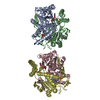
| ||||||||
|---|---|---|---|---|---|---|---|---|---|
| 1 | 
| ||||||||
| 2 | 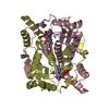
| ||||||||
| Unit cell |
|
-Components
| #1: Protein | Mass: 23937.182 Da / Num. of mol.: 4 Source method: isolated from a genetically manipulated source Source: (gene. exp.) #2: Chemical | ChemComp-FMN /   Mass: 456.344 Da / Num. of mol.: 4 / Source method: obtained synthetically / Formula: C17H21N4O9P Mass: 456.344 Da / Num. of mol.: 4 / Source method: obtained synthetically / Formula: C17H21N4O9P#3: Chemical | ChemComp-NIO /   Mass: 123.109 Da / Num. of mol.: 4 / Source method: obtained synthetically / Formula: C6H5NO2 Mass: 123.109 Da / Num. of mol.: 4 / Source method: obtained synthetically / Formula: C6H5NO2#4: Water | ChemComp-HOH / |  Mass: 18.015 Da / Num. of mol.: 139 / Source method: isolated from a natural source / Formula: H2O Mass: 18.015 Da / Num. of mol.: 139 / Source method: isolated from a natural source / Formula: H2O |
|---|
-Experimental details
-Experiment
| Experiment | Method: X-RAY DIFFRACTION / Number of used crystals: 1 |
|---|
- Sample preparation
| Crystal | Density Matthews: 2.46 Å3/Da / Density % sol: 50.04 % | ||||||||||||||||||||||||||||||
|---|---|---|---|---|---|---|---|---|---|---|---|---|---|---|---|---|---|---|---|---|---|---|---|---|---|---|---|---|---|---|---|
| Crystal grow | Temperature: 291 K / Method: vapor diffusion, hanging drop / pH: 4.6 Details: PEG8K , pH 4.6, VAPOR DIFFUSION, HANGING DROP, temperature 291K | ||||||||||||||||||||||||||||||
| Crystal grow | *PLUS Temperature: 18 ℃ | ||||||||||||||||||||||||||||||
| Components of the solutions | *PLUS
|
-Data collection
| Diffraction | Mean temperature: 100 K |
|---|---|
| Diffraction source | Source: SYNCHROTRON / Site: ESRF  / Beamline: ID14-1 / Wavelength: 0.934 Å / Beamline: ID14-1 / Wavelength: 0.934 Å |
| Detector | Type: MARRESEARCH / Detector: CCD |
| Radiation | Protocol: SINGLE WAVELENGTH / Monochromatic (M) / Laue (L): M / Scattering type: x-ray |
| Radiation wavelength | Wavelength: 0.934 Å / Relative weight: 1 |
| Reflection | Resolution: 1.8→29.91 Å / Num. all: 611309 / Num. obs: 85582 / % possible obs: 98.9 % / Observed criterion σ(F): 0 / Observed criterion σ(I): 0 / Redundancy: 7.1 % / Biso Wilson estimate: 40.2 Å2 / Rmerge(I) obs: 0.067 / Rsym value: 6.7 / Net I/σ(I): 8.4 |
| Reflection shell | Resolution: 1.8→1.86 Å / Redundancy: 6.1 % / Rmerge(I) obs: 0.549 / Mean I/σ(I) obs: 1.3 / Rsym value: 54.9 / % possible all: 97 |
| Reflection | *PLUS Num. measured all: 611309 |
- Processing
| Software |
| ||||||||||||||||||||
|---|---|---|---|---|---|---|---|---|---|---|---|---|---|---|---|---|---|---|---|---|---|
| Refinement | Method to determine structure: MOLECULAR REPLACEMENT Starting model: 1ICR Resolution: 1.8→100 Å / Isotropic thermal model: anisotropic / Cross valid method: THROUGHOUT / σ(F): 0 / σ(I): 0 / Stereochemistry target values: Engh & Huber
| ||||||||||||||||||||
| Displacement parameters | Biso mean: 45 Å2
| ||||||||||||||||||||
| Refine analyze |
| ||||||||||||||||||||
| Refinement step | Cycle: LAST / Resolution: 1.8→100 Å
| ||||||||||||||||||||
| Refine LS restraints |
| ||||||||||||||||||||
| Software | *PLUS Name: CNS / Classification: refinement | ||||||||||||||||||||
| Refinement | *PLUS Highest resolution: 1.8 Å / Lowest resolution: 100 Å / σ(F): 0 / % reflection Rfree: 5 % / Rfactor obs: 0.226 | ||||||||||||||||||||
| Solvent computation | *PLUS | ||||||||||||||||||||
| Displacement parameters | *PLUS Biso mean: 45 Å2 |
