 Movie
Movie Controller

[English] 日本語
 Yorodumi
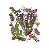
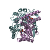
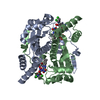
- PDB-1oo5: Studies on the Nitroreductase Prodrug-Activating System. Crystal ... -
+ Open data

- Basic information
| Entry | Database: PDB / ID: 1oo5 | ||||||
|---|---|---|---|---|---|---|---|
| Title | Studies on the Nitroreductase Prodrug-Activating System. Crystal Structures of the Enzyme Active Form and Complexes with the Inhibitor Dicoumarol and Dinitrobenzamide Prodrugs | ||||||
 Components Components | Oxygen-insensitive NAD(P)H nitroreductase | ||||||
 Keywords Keywords | OXIDOREDUCTASE / NAD(P)H-QUINONE REDUCTASE / FMN / NITROREDUCTASE / FLAVOPROTEIN | ||||||
| Function / homology |  Function and homology information Function and homology information6,7-dihydropteridine reductase / 6,7-dihydropteridine reductase activity / NAD(P)H dehydrogenase (quinone) activity / Oxidoreductases / FMN binding / oxidoreductase activity / protein homodimerization activity / membrane / identical protein binding / cytosol Similarity search - Function | ||||||
| Biological species |  | ||||||
| Method |  X-RAY DIFFRACTION / SYNCHROTRON / MOLECULAR REPLACEMENT / Resolution: 2.5 Å X-RAY DIFFRACTION / SYNCHROTRON / MOLECULAR REPLACEMENT / Resolution: 2.5 Å | ||||||
 Authors Authors | Johansson, E. / Parkinson, G.N. / Denny, W.A. / Neidle, S. | ||||||
 Citation Citation | Journal: J.Med.Chem. / Year: 2003 Title: Studies on the Nitroreductase Prodrug-Activating System. Crystal Structures of Complexes with the Inhibitor Dicoumarol and Dinitrobenzamide Prodrugs and of the Enzyme Active Form Authors: Johansson, E. / Parkinson, G.N. / Denny, W.A. / Neidle, S. | ||||||
| History |
|
- Structure visualization
| Structure viewer | Molecule: MolmilJmol/JSmol |
|---|
- Downloads & links
-Download
| PDBx/mmCIF format | 1oo5.cif.gz | 97.6 KB | Display | PDBx/mmCIF format |
|---|---|---|---|---|
| PDB format | pdb1oo5.ent.gz | 75.2 KB | Display | PDB format |
| PDBx/mmJSON format | 1oo5.json.gz | Tree view | PDBx/mmJSON format | |
| Others |  Other downloads Other downloads |
-Validation report
| Arichive directory | https://data.pdbj.org/pub/pdb/validation_reports/oo/1oo5ftp://data.pdbj.org/pub/pdb/validation_reports/oo/1oo5 | HTTPS FTP |
|---|
-Related structure data
| Related structure data |  1idtC  1oo6C  1oonC  1ooqC 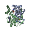 1ds7S C: citing same article ( S: Starting model for refinement |
|---|---|
| Similar structure data |


-Links
 PDBj
PDBj
- Assembly
| Deposited unit | 
| ||||||||
|---|---|---|---|---|---|---|---|---|---|
| 1 |
| ||||||||
| Unit cell |
|
-Components
| #1: Protein | Mass: 23937.182 Da / Num. of mol.: 2 Source method: isolated from a genetically manipulated source Source: (gene. exp.) #2: Chemical |   Mass: 456.344 Da / Num. of mol.: 2 / Source method: obtained synthetically / Formula: C17H21N4O9P Mass: 456.344 Da / Num. of mol.: 2 / Source method: obtained synthetically / Formula: C17H21N4O9P#3: Water | ChemComp-HOH / |  Mass: 18.015 Da / Num. of mol.: 64 / Source method: isolated from a natural source / Formula: H2O Mass: 18.015 Da / Num. of mol.: 64 / Source method: isolated from a natural source / Formula: H2O |
|---|
-Experimental details
-Experiment
| Experiment | Method: X-RAY DIFFRACTION / Number of used crystals: 1 |
|---|
- Sample preparation
| Crystal | Density Matthews: 2.2 Å3/Da / Density % sol: 43.7 % |
|---|---|
| Crystal grow | Temperature: 277 K / Method: vapor diffusion, hanging drop / pH: 4.6 Details: PEG 4000 (25%-29%), 0.1 mM sodium acetate, pH 4.6, VAPOR DIFFUSION, HANGING DROP, temperature 277K |
-Data collection
| Diffraction | Mean temperature: 97 K |
|---|---|
| Diffraction source | Source: SYNCHROTRON / Site: ESRF  / Beamline: ID14-2 / Wavelength: 0.93 Å / Beamline: ID14-2 / Wavelength: 0.93 Å |
| Detector | Type: ADSC QUANTUM 4 / Detector: CCD / Date: Jul 1, 2001 |
| Radiation | Monochromator: diamond (111) germanium (220) / Protocol: SINGLE WAVELENGTH / Monochromatic (M) / Laue (L): M / Scattering type: x-ray |
| Radiation wavelength | Wavelength: 0.93 Å / Relative weight: 1 |
| Reflection | Resolution: 2.5→20 Å / Num. all: 15515 / Num. obs: 15515 / % possible obs: 96.2 % / Observed criterion σ(F): 0 / Observed criterion σ(I): 0 / Biso Wilson estimate: 9.5 Å2 / Rmerge(I) obs: 0.085 / Net I/σ(I): 10 |
| Reflection shell | Highest resolution: 2.5 Å / Rmerge(I) obs: 0.369 / Mean I/σ(I) obs: 1 / Num. unique all: 1402 / % possible all: 89 |
- Processing
| Software |
| ||||||||||||||||||||||||||||||||||||||||||||||||||||||||||||
|---|---|---|---|---|---|---|---|---|---|---|---|---|---|---|---|---|---|---|---|---|---|---|---|---|---|---|---|---|---|---|---|---|---|---|---|---|---|---|---|---|---|---|---|---|---|---|---|---|---|---|---|---|---|---|---|---|---|---|---|---|---|
| Refinement | Method to determine structure: MOLECULAR REPLACEMENT Starting model: PDB ENTRY 1DS7 Resolution: 2.5→19.92 Å / Rfactor Rfree error: 0.009 / Data cutoff high absF: 161733.98 / Data cutoff high rms absF: 161733.98 / Data cutoff low absF: 0 / Isotropic thermal model: RESTRAINED / Cross valid method: THROUGHOUT / σ(F): 2 / Stereochemistry target values: Engh & Huber
| ||||||||||||||||||||||||||||||||||||||||||||||||||||||||||||
| Solvent computation | Solvent model: FLAT MODEL / Bsol: 18.4561 Å2 / ksol: 0.320748 e/Å3 | ||||||||||||||||||||||||||||||||||||||||||||||||||||||||||||
| Displacement parameters | Biso mean: 37.9 Å2
| ||||||||||||||||||||||||||||||||||||||||||||||||||||||||||||
| Refine analyze |
| ||||||||||||||||||||||||||||||||||||||||||||||||||||||||||||
| Refinement step | Cycle: LAST / Resolution: 2.5→19.92 Å
| ||||||||||||||||||||||||||||||||||||||||||||||||||||||||||||
| Refine LS restraints |
| ||||||||||||||||||||||||||||||||||||||||||||||||||||||||||||
| LS refinement shell | Resolution: 2.5→2.66 Å / Rfactor Rfree error: 0.039 / Total num. of bins used: 6
|
