 Movie
Movie Controller

[English] 日本語
 Yorodumi
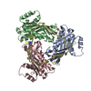
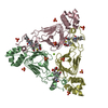
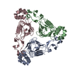
- PDB-1khr: Crystal Structure of Vat(D) in Complex with Virginiamycin and Coe... -
+ Open data

- Basic information
| Entry | Database: PDB / ID: 1khr | ||||||
|---|---|---|---|---|---|---|---|
| Title | Crystal Structure of Vat(D) in Complex with Virginiamycin and Coenzyme A | ||||||
 Components Components | STREPTOGRAMIN A ACETYLTRANSFERASE | ||||||
 Keywords Keywords | transferase/antibiotic / Antibiotic resistance / Acyltransferase / transferase-antibiotic complex | ||||||
| Function / homology |  Function and homology information Function and homology informationacyltransferase activity / Transferases; Acyltransferases; Transferring groups other than aminoacyl groups / response to antibiotic Similarity search - Function | ||||||
| Biological species |  Enterococcus faecium (bacteria) Enterococcus faecium (bacteria) | ||||||
| Method |  X-RAY DIFFRACTION / MOLECULAR REPLACEMENT / Resolution: 2.8 Å X-RAY DIFFRACTION / MOLECULAR REPLACEMENT / Resolution: 2.8 Å | ||||||
 Authors Authors | Sugantino, M. / Roderick, S.L. | ||||||
 Citation Citation | Journal: Biochemistry / Year: 2002 Title: Crystal structure of Vat(D): an acetyltransferase that inactivates streptogramin group A antibiotics. Authors: Sugantino, M. / Roderick, S.L. | ||||||
| History |
|
- Structure visualization
| Structure viewer | Molecule: MolmilJmol/JSmol |
|---|
- Downloads & links
-Download
| PDBx/mmCIF format | 1khr.cif.gz | 255 KB | Display | PDBx/mmCIF format |
|---|---|---|---|---|
| PDB format | pdb1khr.ent.gz | 208.9 KB | Display | PDB format |
| PDBx/mmJSON format | 1khr.json.gz | Tree view | PDBx/mmJSON format | |
| Others |  Other downloads Other downloads |
-Validation report
| Arichive directory | https://data.pdbj.org/pub/pdb/validation_reports/kh/1khrftp://data.pdbj.org/pub/pdb/validation_reports/kh/1khr | HTTPS FTP |
|---|
-Related structure data


-Links
 PDBj
PDBj


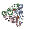
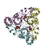
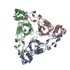
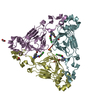
- Assembly
| Deposited unit | 
| ||||||||
|---|---|---|---|---|---|---|---|---|---|
| 1 | 
| ||||||||
| 2 | 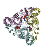
| ||||||||
| Unit cell |
| ||||||||
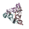
| Details | The biological unit is a trimer. Chains A, B and C form one trimer. Chains D, E and F form one trimer. |
-Components
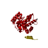
| #1: Protein | Mass: 23675.326 Da / Num. of mol.: 6 Source method: isolated from a genetically manipulated source Source: (gene. exp.) Enterococcus faecium (bacteria) / Gene: satA / Plasmid: pET11a / Species (production host): Escherichia coli / Production host: References: UniProt: P50870, Transferases; Acyltransferases; Transferring groups other than aminoacyl groups #2: Chemical |   Mass: 525.593 Da / Num. of mol.: 3 / Source method: isolated from a natural source / Formula: C28H35N3O7 / Comment: antibiotic*YM Mass: 525.593 Da / Num. of mol.: 3 / Source method: isolated from a natural source / Formula: C28H35N3O7 / Comment: antibiotic*YM#3: Chemical | ChemComp-COA /   Mass: 767.534 Da / Num. of mol.: 6 / Source method: obtained synthetically / Formula: C21H36N7O16P3S Mass: 767.534 Da / Num. of mol.: 6 / Source method: obtained synthetically / Formula: C21H36N7O16P3S#4: Water | ChemComp-HOH / |  Mass: 18.015 Da / Num. of mol.: 112 / Source method: isolated from a natural source / Formula: H2O Mass: 18.015 Da / Num. of mol.: 112 / Source method: isolated from a natural source / Formula: H2O |
|---|
-Experimental details
-Experiment
| Experiment | Method: X-RAY DIFFRACTION / Number of used crystals: 1 |
|---|
- Sample preparation
| Crystal | Density Matthews: 2.84 Å3/Da / Density % sol: 56.74 % | ||||||||||||||||||||||||
|---|---|---|---|---|---|---|---|---|---|---|---|---|---|---|---|---|---|---|---|---|---|---|---|---|---|
| Crystal grow | Temperature: 298 K / Method: vapor diffusion, hanging drop / pH: 8 Details: PEG 400, ethanol, sodium citrate, tris-HCl, pH 8.0, VAPOR DIFFUSION, HANGING DROP, temperature 298K | ||||||||||||||||||||||||
| Crystal grow | *PLUS | ||||||||||||||||||||||||
| Components of the solutions | *PLUS
|
-Data collection
| Diffraction | Mean temperature: 298 K |
|---|---|
| Diffraction source | Source: ROTATING ANODE / Type: RIGAKU RU200 / Wavelength: 1.5418 Å |
| Detector | Type: XENTRONICS / Detector: AREA DETECTOR / Date: Oct 5, 1999 |
| Radiation | Monochromator: GRAPHITE / Protocol: SINGLE WAVELENGTH / Monochromatic (M) / Laue (L): M / Scattering type: x-ray |
| Radiation wavelength | Wavelength: 1.5418 Å / Relative weight: 1 |
| Reflection | Resolution: 2.8→99 Å / Num. all: 37772 / Num. obs: 37772 / % possible obs: 93.5 % / Observed criterion σ(F): 0 / Observed criterion σ(I): 0 / Redundancy: 3.3 % / Biso Wilson estimate: 41.5 Å2 / Rmerge(I) obs: 0.089 |
| Reflection shell | Resolution: 2.8→2.9 Å / Rmerge(I) obs: 0.296 / % possible all: 91.9 |
| Reflection | *PLUS Redundancy: 3.6 % / Num. measured all: 135079 |
| Reflection shell | *PLUS Highest resolution: 2.8 Å / % possible obs: 91.9 % |
- Processing
| Software |
| ||||||||||||||||||||||||||||||||||||||||||||||||||||||||||||||||||||||||||||||||
|---|---|---|---|---|---|---|---|---|---|---|---|---|---|---|---|---|---|---|---|---|---|---|---|---|---|---|---|---|---|---|---|---|---|---|---|---|---|---|---|---|---|---|---|---|---|---|---|---|---|---|---|---|---|---|---|---|---|---|---|---|---|---|---|---|---|---|---|---|---|---|---|---|---|---|---|---|---|---|---|---|---|
| Refinement | Method to determine structure: MOLECULAR REPLACEMENT / Resolution: 2.8→99 Å / Rfactor Rfree error: 0.006 / Data cutoff high absF: 10000000 / Data cutoff low absF: 0.001 / Isotropic thermal model: RESTRAINED / Cross valid method: THROUGHOUT / σ(F): 0 / Stereochemistry target values: Engh & Huber
| ||||||||||||||||||||||||||||||||||||||||||||||||||||||||||||||||||||||||||||||||
| Displacement parameters | Biso mean: 21.2 Å2
| ||||||||||||||||||||||||||||||||||||||||||||||||||||||||||||||||||||||||||||||||
| Refine analyze |
| ||||||||||||||||||||||||||||||||||||||||||||||||||||||||||||||||||||||||||||||||
| Refinement step | Cycle: LAST / Resolution: 2.8→99 Å
| ||||||||||||||||||||||||||||||||||||||||||||||||||||||||||||||||||||||||||||||||
| Refine LS restraints |
| ||||||||||||||||||||||||||||||||||||||||||||||||||||||||||||||||||||||||||||||||
| LS refinement shell | Resolution: 2.8→2.98 Å / Rfactor Rfree error: 0.02 / Total num. of bins used: 6
| ||||||||||||||||||||||||||||||||||||||||||||||||||||||||||||||||||||||||||||||||
| Xplor file |
| ||||||||||||||||||||||||||||||||||||||||||||||||||||||||||||||||||||||||||||||||
| Software | *PLUS Name: X-PLOR(ONLINE) / Version: 3.851 / Classification: refinement | ||||||||||||||||||||||||||||||||||||||||||||||||||||||||||||||||||||||||||||||||
| Refinement | *PLUS Highest resolution: 2.8 Å / σ(F): 0 / % reflection Rfree: 5 % | ||||||||||||||||||||||||||||||||||||||||||||||||||||||||||||||||||||||||||||||||
| Solvent computation | *PLUS | ||||||||||||||||||||||||||||||||||||||||||||||||||||||||||||||||||||||||||||||||
| Displacement parameters | *PLUS Biso mean: 21.2 Å2 | ||||||||||||||||||||||||||||||||||||||||||||||||||||||||||||||||||||||||||||||||
| Refine LS restraints | *PLUS
| ||||||||||||||||||||||||||||||||||||||||||||||||||||||||||||||||||||||||||||||||
| LS refinement shell | *PLUS Highest resolution: 2.8 Å / Rfactor Rfree: 0.321 / % reflection Rfree: 4.8 % / Rfactor Rwork: 0.248 / Rfactor obs: 0.248 |
