 Movie
Movie Controller

[English] 日本語
 Yorodumi
- PDB-3boy: Crystal structure of the HutP antitermination complex bound to th... -
+ Open data

- Basic information
| Entry | Database: PDB / ID: 3boy | |||||||||
|---|---|---|---|---|---|---|---|---|---|---|
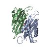
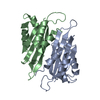
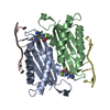
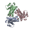
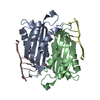
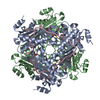
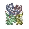
| Title | Crystal structure of the HutP antitermination complex bound to the HUT mRNA | |||||||||
 Components Components |
| |||||||||
 Keywords Keywords | TRANSCRIPTION/RNA / HutP / RNA-binding / HutP-RNA Complex / Anti-termination / Transcription regulation / Activator / Histidine metabolism / TRANSCRIPTION-RNA COMPLEX / Structural Genomics / NPPSFA / National Project on Protein Structural and Functional Analyses / RIKEN Structural Genomics/Proteomics Initiative / RSGI | |||||||||
| Function / homology |  Function and homology information Function and homology information | |||||||||
| Biological species |  | |||||||||
| Method |  X-RAY DIFFRACTION / SYNCHROTRON / MOLECULAR REPLACEMENT / Resolution: 1.7 Å X-RAY DIFFRACTION / SYNCHROTRON / MOLECULAR REPLACEMENT / Resolution: 1.7 Å | |||||||||
 Authors Authors | Kumarevel, T.S. / Balasundaresan, D. / Jeyakanthan, J. / Shinkai, A. / Yokoyama, S. / Kumar, P.K.R. / RIKEN Structural Genomics/Proteomics Initiative (RSGI) | |||||||||
 Citation Citation | Journal: To be Published Title: Crystal Structure of HutP complexed with the 55-mer RNA Authors: Kumarevel, T. / Balasundaresan, D. / Jeyakanthan, J. / Shinkai, A. / Yokoyama, S. / Kumar, P.K.R. #1: Journal: Structure / Year: 2004Title: Crystal Structure of Activated HutP: An RNA Binding Protein that Regulates Transcription of the hut Operon in Bacillus subtilis Authors: Kumarevel, T. / Fujimoto, Z. / Karthe, P. / Oda, M. / Mizuno, H. / Kumar, P.K.R. #2: Journal: NUCLEIC ACIDS RES. / Year: 2004 Title: Identification of important chemical groups of the hut mRNA for HutP interactions that regulate the hut operon in Bacillus subtilis Authors: Kumarevel, T. / Gopinath, S.C.B. / Nishikawa, S. / Mizuno, H. / Kumar, P.K.R. #3: Journal: Nature / Year: 2005Title: Structural basis of HutP-mediated anti-termination and roles of the Mg2+ ion and L-histidine ligand Authors: Kumarevel, T. / Mizuno, H. / Kumar, P.K.R. #4: Journal: Nucleic Acids Res. / Year: 2005Title: Characterization of the metal ion binding site in the anti-terminator protein, HutP, of Bacillus subtilis Authors: Kumarevel, T. / Mizuno, H. / Kumar, P.K.R. | |||||||||
| History |
|
- Structure visualization
| Structure viewer | Molecule: MolmilJmol/JSmol |
|---|
- Downloads & links
-Download
| PDBx/mmCIF format | 3boy.cif.gz | 121.8 KB | Display | PDBx/mmCIF format |
|---|---|---|---|---|
| PDB format | pdb3boy.ent.gz | 91.9 KB | Display | PDB format |
| PDBx/mmJSON format | 3boy.json.gz | Tree view | PDBx/mmJSON format | |
| Others |  Other downloads Other downloads |
-Validation report
| Arichive directory | https://data.pdbj.org/pub/pdb/validation_reports/bo/3boyftp://data.pdbj.org/pub/pdb/validation_reports/bo/3boy | HTTPS FTP |
|---|
-Related structure data
| Related structure data | 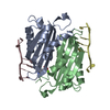 1wmqS S: Starting model for refinement |
|---|---|
| Similar structure data | |
| Other databases |


-Links
 PDBj
PDBj


- Assembly
| Deposited unit | 
| ||||||||
|---|---|---|---|---|---|---|---|---|---|
| 1 | 
| ||||||||
| Unit cell |
|
-Components
| #1: RNA chain | Mass: 6876.931 Da / Num. of mol.: 1 / Source method: obtained synthetically | ||||||
|---|---|---|---|---|---|---|---|
| #2: Protein | Mass: 16101.357 Da / Num. of mol.: 3 / Mutation: V51I Source method: isolated from a genetically manipulated source Source: (gene. exp.) #3: Chemical |   Mass: 24.305 Da / Num. of mol.: 3 / Source method: obtained synthetically / Formula: Mg Mass: 24.305 Da / Num. of mol.: 3 / Source method: obtained synthetically / Formula: Mg#4: Chemical |   Type: L-peptide linking / Mass: 156.162 Da / Num. of mol.: 3 / Source method: obtained synthetically / Formula: C6H10N3O2 Type: L-peptide linking / Mass: 156.162 Da / Num. of mol.: 3 / Source method: obtained synthetically / Formula: C6H10N3O2#5: Water | ChemComp-HOH / |  Mass: 18.015 Da / Num. of mol.: 406 / Source method: isolated from a natural source / Formula: H2O Mass: 18.015 Da / Num. of mol.: 406 / Source method: isolated from a natural source / Formula: H2O |
-Experimental details
-Experiment
| Experiment | Method: X-RAY DIFFRACTION / Number of used crystals: 1 |
|---|
- Sample preparation
| Crystal | Density Matthews: 2.05 Å3/Da / Density % sol: 39.96 % | ||||||||||||||||||||||||||||||||||||
|---|---|---|---|---|---|---|---|---|---|---|---|---|---|---|---|---|---|---|---|---|---|---|---|---|---|---|---|---|---|---|---|---|---|---|---|---|---|
| Crystal grow | Temperature: 293 K / Method: vapor diffusion, hanging drop / pH: 7.4 Details: 40% MPD, 0.1M HEPES, 0.1M MGCL2, pH 7.40, VAPOR DIFFUSION, HANGING DROP, temperature 293K | ||||||||||||||||||||||||||||||||||||
| Components of the solutions |
|
-Data collection
| Diffraction | Mean temperature: 100 K |
|---|---|
| Diffraction source | Source: SYNCHROTRON / Site: SPring-8  / Beamline: BL26B1 / Wavelength: 1 Å / Beamline: BL26B1 / Wavelength: 1 Å |
| Detector | Type: ADSC QUANTUM 4 / Detector: CCD / Date: Apr 18, 2006 |
| Radiation | Monochromator: SI II Channel / Protocol: SINGLE WAVELENGTH / Monochromatic (M) / Laue (L): M / Scattering type: x-ray |
| Radiation wavelength | Wavelength: 1 Å / Relative weight: 1 |
| Reflection | Resolution: 1.7→40 Å / Num. all: 48341 / Num. obs: 48341 / % possible obs: 99.4 % / Observed criterion σ(F): 0 / Observed criterion σ(I): 0 / Redundancy: 3.6 % / Rmerge(I) obs: 0.045 |
| Reflection shell | Resolution: 1.7→1.76 Å / Redundancy: 3.3 % / Rmerge(I) obs: 0.362 / Num. unique all: 4735 / % possible all: 98.4 |
- Processing
| Software |
| ||||||||||||||||||||||||||||||||||||||||||||||||||||||||||||||||||||||||||||||||||||||||||
|---|---|---|---|---|---|---|---|---|---|---|---|---|---|---|---|---|---|---|---|---|---|---|---|---|---|---|---|---|---|---|---|---|---|---|---|---|---|---|---|---|---|---|---|---|---|---|---|---|---|---|---|---|---|---|---|---|---|---|---|---|---|---|---|---|---|---|---|---|---|---|---|---|---|---|---|---|---|---|---|---|---|---|---|---|---|---|---|---|---|---|---|
| Refinement | Method to determine structure: MOLECULAR REPLACEMENT Starting model: 1WMQ Resolution: 1.7→38.24 Å / Cor.coef. Fo:Fc: 0.958 / Cor.coef. Fo:Fc free: 0.936 / SU B: 2.627 / SU ML: 0.088 / Cross valid method: THROUGHOUT / σ(F): 0 / ESU R: 0.131 / ESU R Free: 0.13 / Stereochemistry target values: MAXIMUM LIKELIHOOD
| ||||||||||||||||||||||||||||||||||||||||||||||||||||||||||||||||||||||||||||||||||||||||||
| Solvent computation | Ion probe radii: 0.8 Å / Shrinkage radii: 0.8 Å / VDW probe radii: 1.2 Å / Solvent model: MASK | ||||||||||||||||||||||||||||||||||||||||||||||||||||||||||||||||||||||||||||||||||||||||||
| Displacement parameters | Biso mean: 28.961 Å2
| ||||||||||||||||||||||||||||||||||||||||||||||||||||||||||||||||||||||||||||||||||||||||||
| Refinement step | Cycle: LAST / Resolution: 1.7→38.24 Å
| ||||||||||||||||||||||||||||||||||||||||||||||||||||||||||||||||||||||||||||||||||||||||||
| Refine LS restraints |
| ||||||||||||||||||||||||||||||||||||||||||||||||||||||||||||||||||||||||||||||||||||||||||
| LS refinement shell | Resolution: 1.703→1.747 Å / Total num. of bins used: 20
|
