 Movie
Movie Controller

[English] 日本語
 Yorodumi
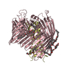
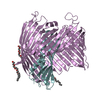
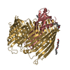
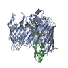
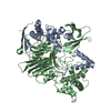
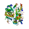
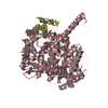
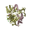
- PDB-5iva: The LPS Transporter LptDE from Pseudomonas aeruginosa, core complex -
+ Open data

- Basic information
| Entry | Database: PDB / ID: 5iva | ||||||
|---|---|---|---|---|---|---|---|
| Title | The LPS Transporter LptDE from Pseudomonas aeruginosa, core complex | ||||||
 Components Components |
| ||||||
 Keywords Keywords | TRANSPORT PROTEIN / LptD / LptE / lipopolysaccharide / Transporter | ||||||
| Function / homology |  Function and homology information Function and homology informationregulation of polysaccharide biosynthetic process / transporter complex / lipopolysaccharide transport / Gram-negative-bacterium-type cell outer membrane assembly / cell outer membrane / lipopolysaccharide binding Similarity search - Function | ||||||
| Biological species |   Pseudomonas aeruginosa (bacteria) Pseudomonas aeruginosa (bacteria) | ||||||
| Method |  X-RAY DIFFRACTION / SYNCHROTRON / MOLECULAR REPLACEMENT / Resolution: 2.988 Å X-RAY DIFFRACTION / SYNCHROTRON / MOLECULAR REPLACEMENT / Resolution: 2.988 Å | ||||||
 Authors Authors | Botos, I. / Buchanan, S.K. | ||||||
 Citation Citation | Journal: Structure / Year: 2016 Title: Structural and Functional Characterization of the LPS Transporter LptDE from Gram-Negative Pathogens. Authors: Botos, I. / Majdalani, N. / Mayclin, S.J. / McCarthy, J.G. / Lundquist, K. / Wojtowicz, D. / Barnard, T.J. / Gumbart, J.C. / Buchanan, S.K. | ||||||
| History |
|
- Structure visualization
| Structure viewer | Molecule: MolmilJmol/JSmol |
|---|
- Downloads & links
-Download
| PDBx/mmCIF format | 5iva.cif.gz | 165.1 KB | Display | PDBx/mmCIF format |
|---|---|---|---|---|
| PDB format | pdb5iva.ent.gz | 130.2 KB | Display | PDB format |
| PDBx/mmJSON format | 5iva.json.gz | Tree view | PDBx/mmJSON format | |
| Others |  Other downloads Other downloads |
-Validation report
| Arichive directory | https://data.pdbj.org/pub/pdb/validation_reports/iv/5ivaftp://data.pdbj.org/pub/pdb/validation_reports/iv/5iva | HTTPS FTP |
|---|
-Related structure data
| Related structure data |  5iv8C  5iv9C 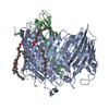 5ixmC  4q35S C: citing same article ( S: Starting model for refinement |
|---|---|
| Similar structure data |

-Links
 PDBj
PDBj


- Assembly
| Deposited unit | 
| ||||||||
|---|---|---|---|---|---|---|---|---|---|
| 1 |
| ||||||||
| Unit cell |
|
-Components
| #1: Protein | Mass: 75433.367 Da / Num. of mol.: 1 / Fragment: unp residues 300-924 Source method: isolated from a genetically manipulated source Source: (gene. exp.) Pseudomonas aeruginosa (bacteria) / Strain: ATCC 15692 / PAO1 / 1C / PRS 101 / LMG 12228 / Gene: lptD, imp, ostA, PA0595 / Production host: | ||
|---|---|---|---|
| #2: Protein | Mass: 21316.590 Da / Num. of mol.: 1 / Fragment: unp residues 21-207 Source method: isolated from a genetically manipulated source Source: (gene. exp.) Pseudomonas aeruginosa (bacteria) / Gene: lptE, PAMH19_1038 / Production host: | ||
| #3: Chemical | ChemComp-C8E / (   Mass: 306.438 Da / Num. of mol.: 7 / Source method: obtained synthetically / Formula: C16H34O5 / Comment: C8E, detergent*YM Mass: 306.438 Da / Num. of mol.: 7 / Source method: obtained synthetically / Formula: C16H34O5 / Comment: C8E, detergent*YM#4: Water | ChemComp-HOH / |  Mass: 18.015 Da / Num. of mol.: 4 / Source method: isolated from a natural source / Formula: H2O Mass: 18.015 Da / Num. of mol.: 4 / Source method: isolated from a natural source / Formula: H2O |
-Experimental details
-Experiment
| Experiment | Method: X-RAY DIFFRACTION / Number of used crystals: 1 |
|---|
- Sample preparation
| Crystal | Density Matthews: 3.47 Å3/Da / Density % sol: 64.59 % |
|---|---|
| Crystal grow | Temperature: 294 K / Method: vapor diffusion, hanging drop / pH: 5.5 Details: 100 mM sodium citrate pH 5.5, 100 mM sodium chloride, 100 mM lithium chloride, 12% PEG 4000 |
-Data collection
| Diffraction | Mean temperature: 90 K |
|---|---|
| Diffraction source | Source: SYNCHROTRON / Site: APS  / Beamline: 23-ID-B / Wavelength: 1 Å / Beamline: 23-ID-B / Wavelength: 1 Å |
| Detector | Type: MARMOSAIC 300 mm CCD / Detector: CCD / Date: Nov 22, 2013 |
| Radiation | Protocol: SINGLE WAVELENGTH / Monochromatic (M) / Laue (L): M / Scattering type: x-ray |
| Radiation wavelength | Wavelength: 1 Å / Relative weight: 1 |
| Reflection | Resolution: 2.988→49.089 Å / Num. obs: 27493 / % possible obs: 100 % / Redundancy: 7.2 % / Net I/σ(I): 12.4 |
- Processing
| Software |
| |||||||||||||||||||||||||||||||||||||||||||||||||||||||||||||||||||||||||||||
|---|---|---|---|---|---|---|---|---|---|---|---|---|---|---|---|---|---|---|---|---|---|---|---|---|---|---|---|---|---|---|---|---|---|---|---|---|---|---|---|---|---|---|---|---|---|---|---|---|---|---|---|---|---|---|---|---|---|---|---|---|---|---|---|---|---|---|---|---|---|---|---|---|---|---|---|---|---|---|
| Refinement | Method to determine structure: MOLECULAR REPLACEMENT Starting model: 4Q35 Resolution: 2.988→49.089 Å / SU ML: 0.47 / Cross valid method: FREE R-VALUE / σ(F): 1.34 / Phase error: 31.3 / Stereochemistry target values: ML
| |||||||||||||||||||||||||||||||||||||||||||||||||||||||||||||||||||||||||||||
| Solvent computation | Shrinkage radii: 0.9 Å / VDW probe radii: 1.11 Å / Solvent model: FLAT BULK SOLVENT MODEL | |||||||||||||||||||||||||||||||||||||||||||||||||||||||||||||||||||||||||||||
| Refinement step | Cycle: LAST / Resolution: 2.988→49.089 Å
| |||||||||||||||||||||||||||||||||||||||||||||||||||||||||||||||||||||||||||||
| Refine LS restraints |
| |||||||||||||||||||||||||||||||||||||||||||||||||||||||||||||||||||||||||||||
| LS refinement shell |
|
