 Movie
Movie Controller

[English] 日本語
 Yorodumi
- PDB-1kgq: Crystal Structure of Tetrahydrodipicolinate N-Succinyltransferase... -
+ Open data

- Basic information
| Entry | Database: PDB / ID: 1kgq | ||||||
|---|---|---|---|---|---|---|---|
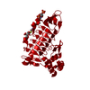
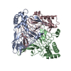
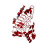
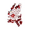
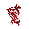
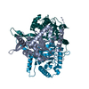
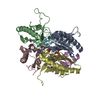
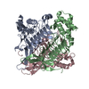
| Title | Crystal Structure of Tetrahydrodipicolinate N-Succinyltransferase in Complex with L-2-aminopimelate and Succinamide-CoA | ||||||
 Components Components | 2,3,4,5-TETRAHYDROPYRIDINE-2-CARBOXYLATE N-SUCCINYLTRANSFERASE | ||||||
 Keywords Keywords | TRANSFERASE / LEFT-HANDED PARALLEL BETA HELIX | ||||||
| Function / homology |  Function and homology information Function and homology information2,3,4,5-tetrahydropyridine-2,6-dicarboxylate N-succinyltransferase / 2,3,4,5-tetrahydropyridine-2,6-dicarboxylate N-succinyltransferase activity / : / : / nucleotidyltransferase activity / cytoplasm Similarity search - Function | ||||||
| Biological species |  Mycobacterium bovis (bacteria) Mycobacterium bovis (bacteria) | ||||||
| Method |  X-RAY DIFFRACTION / MOLECULAR REPLACEMENT / Resolution: 2 Å X-RAY DIFFRACTION / MOLECULAR REPLACEMENT / Resolution: 2 Å | ||||||
 Authors Authors | Beaman, T.W. / Vogel, K.W. / Drueckhammer, D.G. / Blanchard, J.S. / Roderick, S.L. | ||||||
 Citation Citation | Journal: Protein Sci. / Year: 2002 Title: Acyl group specificity at the active site of tetrahydridipicolinate N-succinyltransferase. Authors: Beaman, T.W. / Vogel, K.W. / Drueckhammer, D.G. / Blanchard, J.S. / Roderick, S.L. | ||||||
| History |
|
- Structure visualization
| Structure viewer | Molecule: MolmilJmol/JSmol |
|---|
- Downloads & links
-Download
| PDBx/mmCIF format | 1kgq.cif.gz | 69.8 KB | Display | PDBx/mmCIF format |
|---|---|---|---|---|
| PDB format | pdb1kgq.ent.gz | 50.8 KB | Display | PDB format |
| PDBx/mmJSON format | 1kgq.json.gz | Tree view | PDBx/mmJSON format | |
| Others |  Other downloads Other downloads |
-Validation report
| Arichive directory | https://data.pdbj.org/pub/pdb/validation_reports/kg/1kgqftp://data.pdbj.org/pub/pdb/validation_reports/kg/1kgq | HTTPS FTP |
|---|
-Related structure data


-Links
 PDBj
PDBj


- Assembly
| Deposited unit | 
| ||||||||
|---|---|---|---|---|---|---|---|---|---|
| 1 | 
| ||||||||
| Unit cell |
| ||||||||
| Details | The biological assembly is a trimer generated from the monomer in the asymmetric unit by the operations: 1-y,x-y,z and -x+y,1-x,z |
-Components
| #1: Protein | Mass: 29918.990 Da / Num. of mol.: 1 Source method: isolated from a genetically manipulated source Source: (gene. exp.) Mycobacterium bovis (bacteria) / Gene: dapD / Plasmid: pET3a / Species (production host): Escherichia coli / Production host: References: UniProt: P56220, 2,3,4,5-tetrahydropyridine-2,6-dicarboxylate N-succinyltransferase |
|---|---|
| #2: Chemical | ChemComp-NPI / (  Type: L-peptide linking / Mass: 175.182 Da / Num. of mol.: 1 / Source method: obtained synthetically / Formula: C7H13NO4 Type: L-peptide linking / Mass: 175.182 Da / Num. of mol.: 1 / Source method: obtained synthetically / Formula: C7H13NO4 |
| #3: Chemical | ChemComp-SCO /   Mass: 850.557 Da / Num. of mol.: 1 / Source method: obtained synthetically / Formula: C25H41N8O19P3 Mass: 850.557 Da / Num. of mol.: 1 / Source method: obtained synthetically / Formula: C25H41N8O19P3 |
| #4: Water | ChemComp-HOH /  Mass: 18.015 Da / Num. of mol.: 85 / Source method: isolated from a natural source / Formula: H2O Mass: 18.015 Da / Num. of mol.: 85 / Source method: isolated from a natural source / Formula: H2O |
-Experimental details
-Experiment
| Experiment | Method: X-RAY DIFFRACTION / Number of used crystals: 1 |
|---|
- Sample preparation
| Crystal | Density Matthews: 2.15 Å3/Da / Density % sol: 42.66 % | ||||||||||||||||||||||||||||||||||||||||||||||||
|---|---|---|---|---|---|---|---|---|---|---|---|---|---|---|---|---|---|---|---|---|---|---|---|---|---|---|---|---|---|---|---|---|---|---|---|---|---|---|---|---|---|---|---|---|---|---|---|---|---|
| Crystal grow | Temperature: 298 K / Method: vapor diffusion, hanging drop / pH: 6.4 Details: PEG 4000, ammonium sulfate, MES, pH 6.4, VAPOR DIFFUSION, HANGING DROP, temperature 298K | ||||||||||||||||||||||||||||||||||||||||||||||||
| Crystal grow | *PLUS pH: 7.5 / Details: Binder, D.A., (1996) Proteins, 26, 115. | ||||||||||||||||||||||||||||||||||||||||||||||||
| Components of the solutions | *PLUS
|
-Data collection
| Diffraction | Mean temperature: 298 K |
|---|---|
| Diffraction source | Source: ROTATING ANODE / Type: RIGAKU RU200 / Wavelength: 1.5418 Å |
| Detector | Type: XENTRONICS / Detector: AREA DETECTOR / Date: Apr 26, 1999 |
| Radiation | Monochromator: GRAPHITE / Protocol: SINGLE WAVELENGTH / Monochromatic (M) / Laue (L): M / Scattering type: x-ray |
| Radiation wavelength | Wavelength: 1.5418 Å / Relative weight: 1 |
| Reflection | Resolution: 2→99 Å / Num. all: 16693 / Num. obs: 16693 / % possible obs: 99.5 % / Observed criterion σ(F): 0 / Observed criterion σ(I): 0 / Rmerge(I) obs: 0.048 |
| Reflection shell | Resolution: 2→2.1 Å / Rmerge(I) obs: 0.217 / % possible all: 97.9 |
| Reflection | *PLUS Highest resolution: 2 Å / Redundancy: 3.5 % / Num. measured all: 59293 / Rmerge(I) obs: 0.048 |
| Reflection shell | *PLUS % possible obs: 97.9 % / Rmerge(I) obs: 0.217 / Mean I/σ(I) obs: 3.2 |
- Processing
| Software |
| ||||||||||||||||||||
|---|---|---|---|---|---|---|---|---|---|---|---|---|---|---|---|---|---|---|---|---|---|
| Refinement | Method to determine structure: MOLECULAR REPLACEMENT / Resolution: 2→99 Å / Cross valid method: THROUGHOUT / σ(F): 2 / Stereochemistry target values: TNT
| ||||||||||||||||||||
| Refinement step | Cycle: LAST / Resolution: 2→99 Å
| ||||||||||||||||||||
| Refine LS restraints |
| ||||||||||||||||||||
| Refinement | *PLUS % reflection Rfree: 5 % / Rfactor obs: 0.179 / Rfactor Rfree: 0.25 / Rfactor Rwork: 0.174 | ||||||||||||||||||||
| Solvent computation | *PLUS | ||||||||||||||||||||
| Displacement parameters | *PLUS | ||||||||||||||||||||
| LS refinement shell | *PLUS Rfactor Rfree: 0.341 / Rfactor Rwork: 0.27 / Rfactor obs: 0.27 |
