 Movie
Movie Controller

[English] 日本語
 Yorodumi
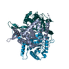
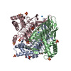
- PDB-1tdt: THREE-DIMENSIONAL STRUCTURE OF TETRAHYDRODIPICOLINATE-N-SUCCINYLT... -
+ Open data

- Basic information
| Entry | Database: PDB / ID: 1tdt | ||||||
|---|---|---|---|---|---|---|---|
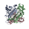
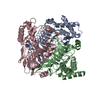
| Title | THREE-DIMENSIONAL STRUCTURE OF TETRAHYDRODIPICOLINATE-N-SUCCINYLTRANSFERASE | ||||||
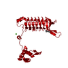
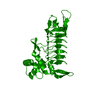
 Components Components | TETRAHYDRODIPICOLINATE-N-SUCCINYLTRANSFERASE | ||||||
 Keywords Keywords | TRANSFERASE / SUCCINYLTRANSFERASE / LYSINE METABOLISM / HEXAPEPTIDE TRANSFERASE / CELL WALL BIOSYNTHESIS | ||||||
| Function / homology |  Function and homology information Function and homology information2,3,4,5-tetrahydropyridine-2,6-dicarboxylate N-succinyltransferase / 2,3,4,5-tetrahydropyridine-2,6-dicarboxylate N-succinyltransferase activity / : / : / nucleotidyltransferase activity / cytoplasm Similarity search - Function | ||||||
| Biological species |  Mycobacterium bovis (bacteria) Mycobacterium bovis (bacteria) | ||||||
| Method |  X-RAY DIFFRACTION / MIR - 2 HEAVY ATOM DERIVATIVES / Resolution: 2.2 Å X-RAY DIFFRACTION / MIR - 2 HEAVY ATOM DERIVATIVES / Resolution: 2.2 Å | ||||||
 Authors Authors | Beaman, T.W. / Binder, D.W. / Blanchard, J.S. / Roderick, S.L. | ||||||
 Citation Citation | Journal: Biochemistry / Year: 1997 Title: Three-dimensional structure of tetrahydrodipicolinate N-succinyltransferase. Authors: Beaman, T.W. / Binder, D.A. / Blanchard, J.S. / Roderick, S.L. #1: Journal: Proteins / Year: 1996Title: Crystallization and Preliminary Crystallographic Analysis of Tetrahydrodipicolinate-N-Succinyltransferase Authors: Binder, D.A. / Blanchard, J.S. / Roderick, S.L. | ||||||
| History |
|
- Structure visualization
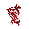
| Structure viewer | Molecule: MolmilJmol/JSmol |
|---|
- Downloads & links
-Download
| PDBx/mmCIF format | 1tdt.cif.gz | 155 KB | Display | PDBx/mmCIF format |
|---|---|---|---|---|
| PDB format | pdb1tdt.ent.gz | 121.9 KB | Display | PDB format |
| PDBx/mmJSON format | 1tdt.json.gz | Tree view | PDBx/mmJSON format | |
| Others |  Other downloads Other downloads |
-Validation report
| Arichive directory | https://data.pdbj.org/pub/pdb/validation_reports/td/1tdtftp://data.pdbj.org/pub/pdb/validation_reports/td/1tdt | HTTPS FTP |
|---|
-Related structure data
| Similar structure data |
|---|

-Links
 PDBj
PDBj


- Assembly
| Deposited unit | 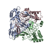
| ||||||||||||
|---|---|---|---|---|---|---|---|---|---|---|---|---|---|
| 1 |
| ||||||||||||
| Unit cell |
| ||||||||||||
| Noncrystallographic symmetry (NCS) | NCS oper:
|
-Components
| #1: Protein | Mass: 28249.055 Da / Num. of mol.: 3 Source method: isolated from a genetically manipulated source Source: (gene. exp.) Mycobacterium bovis (bacteria) / Cell line: BL21 / Plasmid: PET3A / Species (production host): Escherichia coli / Production host: #2: Water | ChemComp-HOH / |  Mass: 18.015 Da / Num. of mol.: 215 / Source method: isolated from a natural source / Formula: H2O Mass: 18.015 Da / Num. of mol.: 215 / Source method: isolated from a natural source / Formula: H2O |
|---|
-Experimental details
-Experiment
| Experiment | Method: X-RAY DIFFRACTION / Number of used crystals: 3 |
|---|
- Sample preparation
| Crystal | Density Matthews: 2.36 Å3/Da / Density % sol: 48 % | |||||||||||||||||||||||||
|---|---|---|---|---|---|---|---|---|---|---|---|---|---|---|---|---|---|---|---|---|---|---|---|---|---|---|
| Crystal grow | pH: 7.5 Details: 14-19 % (W/V) POLYETHYLENEGLYCOL 4000 200 MM AMMONIUM SULFATE 6-12 % 2-PROPANOL 100 MM HEPES, PH 7.5, | |||||||||||||||||||||||||
| Crystal grow | *PLUS Method: unknown | |||||||||||||||||||||||||
| Components of the solutions | *PLUS
|
-Data collection
| Diffraction | Mean temperature: 293 K |
|---|---|
| Diffraction source | Source: ROTATING ANODE / Type: RIGAKU RUH2R / Wavelength: 1.5418 |
| Detector | Type: SIEMENS-NICOLET X100 / Detector: AREA DETECTOR / Date: 1996 |
| Radiation | Monochromator: GRAPHITE(002) / Monochromatic (M) / Laue (L): M / Scattering type: x-ray |
| Radiation wavelength | Wavelength: 1.5418 Å / Relative weight: 1 |
| Reflection | Highest resolution: 2.2 Å / Num. obs: 38447 / % possible obs: 90 % / Observed criterion σ(I): 0 / Redundancy: 3.2 % / Rmerge(I) obs: 0.067 |
| Reflection shell | Resolution: 2.2→2.3 Å / % possible all: 73 |
| Reflection | *PLUS Num. measured all: 137056 |
| Reflection shell | *PLUS % possible obs: 73 % |
- Processing
| Software |
| ||||||||||||||||||||||||||||||||||||||||||||||||||
|---|---|---|---|---|---|---|---|---|---|---|---|---|---|---|---|---|---|---|---|---|---|---|---|---|---|---|---|---|---|---|---|---|---|---|---|---|---|---|---|---|---|---|---|---|---|---|---|---|---|---|---|
| Refinement | Method to determine structure: MIR - 2 HEAVY ATOM DERIVATIVES Highest resolution: 2.2 Å / Isotropic thermal model: BCORREL.DAT (WEIGHTS GIVEN ABOVE) / σ(F): 0 / Stereochemistry target values: PROTGEO.DAT
| ||||||||||||||||||||||||||||||||||||||||||||||||||
| Solvent computation | Bsol: 187.3 Å2 / ksol: 0.679 e/Å3 | ||||||||||||||||||||||||||||||||||||||||||||||||||
| Refinement step | Cycle: LAST / Highest resolution: 2.2 Å
| ||||||||||||||||||||||||||||||||||||||||||||||||||
| Refine LS restraints |
| ||||||||||||||||||||||||||||||||||||||||||||||||||
| Software | *PLUS Name: TNT / Classification: refinement | ||||||||||||||||||||||||||||||||||||||||||||||||||
| Refinement | *PLUS Rfactor obs: 0.17 / Rfactor Rfree: 0.232 / Rfactor Rwork: 0.17 | ||||||||||||||||||||||||||||||||||||||||||||||||||
| Solvent computation | *PLUS | ||||||||||||||||||||||||||||||||||||||||||||||||||
| Displacement parameters | *PLUS | ||||||||||||||||||||||||||||||||||||||||||||||||||
| Refine LS restraints | *PLUS
|
