 Movie
Movie Controller

[English] 日本語
 Yorodumi
- PDB-1jt9: Structure of the mutant F174A T form of the Glucosamine-6-Phospha... -
+ Open data

- Basic information
| Entry | Database: PDB / ID: 1jt9 | ||||||
|---|---|---|---|---|---|---|---|
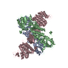
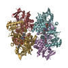
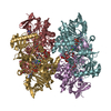
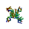
| Title | Structure of the mutant F174A T form of the Glucosamine-6-Phosphate deaminase from E.coli | ||||||
 Components Components | Glucosamine-6-Phosphate deaminase | ||||||
 Keywords Keywords | HYDROLASE / ALLOSTERIC ENZYME / ENTROPIC EFFECTS / ALDOSE-KETOSE ISOMERASE / STRUCTURAL FLEXIBILITY | ||||||
| Function / homology |  Function and homology information Function and homology informationD-glucosamine catabolic process / glucosamine-6-phosphate deaminase / glucosamine-6-phosphate deaminase activity / N-acetylglucosamine catabolic process / N-acetylneuraminate catabolic process / UDP-N-acetylglucosamine biosynthetic process / carbohydrate metabolic process / identical protein binding / cytosol / cytoplasm Similarity search - Function | ||||||
| Biological species |  | ||||||
| Method |  X-RAY DIFFRACTION / SYNCHROTRON / MOLECULAR REPLACEMENT / Resolution: 2.06 Å X-RAY DIFFRACTION / SYNCHROTRON / MOLECULAR REPLACEMENT / Resolution: 2.06 Å | ||||||
 Authors Authors | Bustos-Jaimes, I. / Sosa-Peinado, A. / Rudino-Pinera, E. / Horjales, E. / Calcagno, M.L. | ||||||
 Citation Citation | Journal: J.Mol.Biol. / Year: 2002 Title: On the role of the conformational flexibility of the active-site lid on the allosteric kinetics of glucosamine-6-phosphate deaminase. Authors: Bustos-Jaimes, I. / Sosa-Peinado, A. / Rudino-Pinera, E. / Horjales, E. / Calcagno, M.L. | ||||||
| History |
|
- Structure visualization
| Structure viewer | Molecule: MolmilJmol/JSmol |
|---|
- Downloads & links
-Download
| PDBx/mmCIF format | 1jt9.cif.gz | 71.1 KB | Display | PDBx/mmCIF format |
|---|---|---|---|---|
| PDB format | pdb1jt9.ent.gz | 52.7 KB | Display | PDB format |
| PDBx/mmJSON format | 1jt9.json.gz | Tree view | PDBx/mmJSON format | |
| Others |  Other downloads Other downloads |
-Validation report
| Arichive directory | https://data.pdbj.org/pub/pdb/validation_reports/jt/1jt9ftp://data.pdbj.org/pub/pdb/validation_reports/jt/1jt9 | HTTPS FTP |
|---|
-Related structure data
| Related structure data |  1cd5S S: Starting model for refinement |
|---|---|
| Similar structure data |


-Links
 PDBj
PDBj- Assembly
| Deposited unit | 
| ||||||||
|---|---|---|---|---|---|---|---|---|---|
| 1 | x 6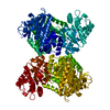
| ||||||||
| 2 | 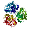
| ||||||||
| Unit cell |
|
-Components
| #1: Protein | Mass: 29736.113 Da / Num. of mol.: 1 / Mutation: F174A Source method: isolated from a genetically manipulated source Source: (gene. exp.) References: UniProt: P0A759, glucosamine-6-phosphate deaminase |
|---|---|
| #2: Water | ChemComp-HOH /  Mass: 18.015 Da / Num. of mol.: 317 / Source method: isolated from a natural source / Formula: H2O Mass: 18.015 Da / Num. of mol.: 317 / Source method: isolated from a natural source / Formula: H2O |
| Has protein modification | Y |
-Experimental details
-Experiment
| Experiment | Method: X-RAY DIFFRACTION / Number of used crystals: 1 |
|---|
- Sample preparation
| Crystal grow | Temperature: 298 K / Method: vapor diffusion, hanging drop / pH: 7 Details: HEPES, sodium acetate, pH 7.0, VAPOR DIFFUSION, HANGING DROP, temperature 298K | ||||||||||||||||||||||||
|---|---|---|---|---|---|---|---|---|---|---|---|---|---|---|---|---|---|---|---|---|---|---|---|---|---|
| Crystal grow | *PLUS Temperature: 18 ℃ | ||||||||||||||||||||||||
| Components of the solutions | *PLUS
|
-Data collection
| Diffraction | Mean temperature: 110 K |
|---|---|
| Diffraction source | Source: SYNCHROTRON / Site: SSRL  / Beamline: BL7-1 / Wavelength: 1.08 Å / Beamline: BL7-1 / Wavelength: 1.08 Å |
| Detector | Type: MARRESEARCH / Detector: IMAGE PLATE / Date: Jan 15, 2000 |
| Radiation | Monochromator: Mirrors / Protocol: SINGLE WAVELENGTH / Monochromatic (M) / Laue (L): M / Scattering type: x-ray |
| Radiation wavelength | Wavelength: 1.08 Å / Relative weight: 1 |
| Reflection | Resolution: 2.02→50 Å / % possible obs: 99 % / Redundancy: 3.4 % / Rsym value: 0.055 / Net I/σ(I): 11.1 |
| Reflection shell | Resolution: 2.06→2.12 Å / Redundancy: 3.2 % / Mean I/σ(I) obs: 2.3 / Rsym value: 0.334 |
| Reflection | *PLUS Lowest resolution: 50 Å / Num. obs: 41650 / % possible obs: 99 % / Rmerge(I) obs: 0.05 |
| Reflection shell | *PLUS Rmerge(I) obs: 0.334 / Mean I/σ(I) obs: 11.1 |
- Processing
| Software |
| |||||||||||||||||||||||||
|---|---|---|---|---|---|---|---|---|---|---|---|---|---|---|---|---|---|---|---|---|---|---|---|---|---|---|
| Refinement | Method to determine structure: MOLECULAR REPLACEMENT Starting model: PDB entry 1cd5 Resolution: 2.06→47.13 Å / Isotropic thermal model: Isotropic / Cross valid method: THROUGHOUT / σ(F): 0 / Stereochemistry target values: Engh & Huber
| |||||||||||||||||||||||||
| Displacement parameters | Biso mean: 36.1 Å2
| |||||||||||||||||||||||||
| Refine analyze | Luzzati coordinate error obs: 0.24 Å / Luzzati d res low obs: 5 Å / Luzzati sigma a obs: 0.19 Å | |||||||||||||||||||||||||
| Refinement step | Cycle: LAST / Resolution: 2.06→47.13 Å
| |||||||||||||||||||||||||
| Refine LS restraints |
| |||||||||||||||||||||||||
| LS refinement shell | Resolution: 2.06→2.2 Å / Rfactor Rfree error: 0.01
| |||||||||||||||||||||||||
| Software | *PLUS Name: CNS / Version: 1 / Classification: refinement | |||||||||||||||||||||||||
| Refinement | *PLUS σ(F): 0 / Rfactor obs: 0.204 | |||||||||||||||||||||||||
| Solvent computation | *PLUS | |||||||||||||||||||||||||
| Displacement parameters | *PLUS Biso mean: 36.1 Å2 | |||||||||||||||||||||||||
| Refine LS restraints | *PLUS
|
