 Movie
Movie Controller

[English] 日本語
 Yorodumi
- PDB-1jlr: STRUCTURE OF THE URACIL PHOSPHORIBOSYLTRANSFERASE GTP COMPLEX 2 M... -
+ Open data

- Basic information
| Entry | Database: PDB / ID: 1jlr | ||||||
|---|---|---|---|---|---|---|---|
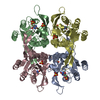
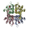
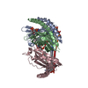
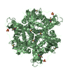
| Title | STRUCTURE OF THE URACIL PHOSPHORIBOSYLTRANSFERASE GTP COMPLEX 2 MUTANT C128V | ||||||
 Components Components | Uracil Phosphoribosyltransferase | ||||||
 Keywords Keywords | TRANSFERASE / GLYCOSYLTRANSFERASE / UPRTASE / GTP activated / tetramer | ||||||
| Function / homology |  Function and homology information Function and homology informationuracil phosphoribosyltransferase / uracil phosphoribosyltransferase activity / UMP salvage / GTP binding Similarity search - Function | ||||||
| Biological species |  | ||||||
| Method |  X-RAY DIFFRACTION / MOLECULAR REPLACEMENT / Resolution: 2.45 Å X-RAY DIFFRACTION / MOLECULAR REPLACEMENT / Resolution: 2.45 Å | ||||||
| Model details | URACIL PHOSPHORIBOSYLTRANSFERASE (E.C.2.4.2.9) | ||||||
 Authors Authors | Schumacher, M.A. / Bashor, C.J. / Otsu, K. / Zu, S. / Parry, R. / Ulmman, B. / Brennan, R.G. | ||||||
 Citation Citation | Journal: Proc.Natl.Acad.Sci.USA / Year: 2002 Title: The structural mechanism of GTP stabilized oligomerization and catalytic activation of the Toxoplasma gondii uracil phosphoribosyltransferase. Authors: Schumacher, M.A. / Bashor, C.J. / Song, M.H. / Otsu, K. / Zhu, S. / Parry, R.J. / Ullman, B. / Brennan, R.G. | ||||||
| History |
|
- Structure visualization
| Structure viewer | Molecule: MolmilJmol/JSmol |
|---|
- Downloads & links
-Download
| PDBx/mmCIF format | 1jlr.cif.gz | 202.8 KB | Display | PDBx/mmCIF format |
|---|---|---|---|---|
| PDB format | pdb1jlr.ent.gz | 162.3 KB | Display | PDB format |
| PDBx/mmJSON format | 1jlr.json.gz | Tree view | PDBx/mmJSON format | |
| Others |  Other downloads Other downloads |
-Validation report
| Arichive directory | https://data.pdbj.org/pub/pdb/validation_reports/jl/1jlrftp://data.pdbj.org/pub/pdb/validation_reports/jl/1jlr | HTTPS FTP |
|---|
-Related structure data
| Related structure data |  1jlsC  1bd3S S: Starting model for refinement C: citing same article ( |
|---|---|
| Similar structure data |


-Links
 PDBj
PDBj
- Assembly
| Deposited unit | 
| ||||||||
|---|---|---|---|---|---|---|---|---|---|
| 1 |
| ||||||||
| Unit cell |
|
-Components
| #1: Protein | Mass: 27558.246 Da / Num. of mol.: 4 / Mutation: C1128V Source method: isolated from a genetically manipulated source Source: (gene. exp.)  References: UniProt: Q26998, uracil phosphoribosyltransferase #2: Chemical | ChemComp-PO4 /   Mass: 94.971 Da / Num. of mol.: 8 / Source method: obtained synthetically / Formula: PO4 Mass: 94.971 Da / Num. of mol.: 8 / Source method: obtained synthetically / Formula: PO4#3: Chemical | ChemComp-GTP /   Mass: 523.180 Da / Num. of mol.: 4 / Source method: obtained synthetically / Formula: C10H16N5O14P3 / Comment: GTP, energy-carrying molecule*YM Mass: 523.180 Da / Num. of mol.: 4 / Source method: obtained synthetically / Formula: C10H16N5O14P3 / Comment: GTP, energy-carrying molecule*YM#4: Water | ChemComp-HOH / |  Mass: 18.015 Da / Num. of mol.: 159 / Source method: isolated from a natural source / Formula: H2O Mass: 18.015 Da / Num. of mol.: 159 / Source method: isolated from a natural source / Formula: H2O |
|---|
-Experimental details
-Experiment
| Experiment | Method: X-RAY DIFFRACTION / Number of used crystals: 1 |
|---|
- Sample preparation
| Crystal | Density Matthews: 2.52 Å3/Da / Density % sol: 51.21 % | ||||||||||||||||||||||||||||
|---|---|---|---|---|---|---|---|---|---|---|---|---|---|---|---|---|---|---|---|---|---|---|---|---|---|---|---|---|---|
| Crystal grow | Temperature: 278 K / Method: vapor diffusion, hanging drop / pH: 5.6 Details: Citrate buffer, NaCl, Ammonium phosphate, pH 5.6, VAPOR DIFFUSION, HANGING DROP, temperature 278K | ||||||||||||||||||||||||||||
| Crystal grow | *PLUS pH: 4.7 | ||||||||||||||||||||||||||||
| Components of the solutions | *PLUS
|
-Data collection
| Diffraction | Mean temperature: 298 K |
|---|---|
| Diffraction source | Source: ROTATING ANODE / Type: RIGAKU RU300 / Wavelength: 1.5418 Å |
| Detector | Type: RIGAKU RAXIS IV / Detector: IMAGE PLATE / Date: Mar 23, 1994 / Details: yale mirrors |
| Radiation | Monochromator: mirrors / Protocol: SINGLE WAVELENGTH / Monochromatic (M) / Laue (L): M / Scattering type: x-ray |
| Radiation wavelength | Wavelength: 1.5418 Å / Relative weight: 1 |
| Reflection | Resolution: 2.45→16.1 Å / Num. obs: 34646 / % possible obs: 86.6 % / Observed criterion σ(F): 0 / Observed criterion σ(I): 0 / Redundancy: 3 % / Biso Wilson estimate: 11.6 Å2 / Rmerge(I) obs: 0.039 / Rsym value: 0.039 / Net I/σ(I): 10.6 |
| Reflection shell | Resolution: 2.45→2.6 Å / Redundancy: 2 % / Rmerge(I) obs: 0.143 / Mean I/σ(I) obs: 3.8 / Num. unique all: 2240 / Rsym value: 0.143 / % possible all: 67 |
| Reflection | *PLUS Num. measured all: 77473 |
| Reflection shell | *PLUS Highest resolution: 2.4 Å |
- Processing
| Software |
| ||||||||||||||||||||
|---|---|---|---|---|---|---|---|---|---|---|---|---|---|---|---|---|---|---|---|---|---|
| Refinement | Method to determine structure: MOLECULAR REPLACEMENT Starting model: 1BD3 Resolution: 2.45→16.14 Å / Rfactor Rfree error: 0.004 / Data cutoff high absF: 522583.02 / Data cutoff low absF: 0 / Isotropic thermal model: RESTRAINED / Cross valid method: THROUGHOUT / σ(F): 0 / Stereochemistry target values: Engh & Huber
| ||||||||||||||||||||
| Solvent computation | Solvent model: FLAT MODEL / Bsol: 29.5739 Å2 / ksol: 0.300439 e/Å3 | ||||||||||||||||||||
| Displacement parameters | Biso mean: 25.1 Å2
| ||||||||||||||||||||
| Refine analyze |
| ||||||||||||||||||||
| Refinement step | Cycle: LAST / Resolution: 2.45→16.14 Å
| ||||||||||||||||||||
| Refine LS restraints |
| ||||||||||||||||||||
| LS refinement shell | Resolution: 2.45→2.6 Å / Rfactor Rfree error: 0.016 / Total num. of bins used: 6
| ||||||||||||||||||||
| Software | *PLUS Name: CNS / Version: 1 / Classification: refinement | ||||||||||||||||||||
| Refinement | *PLUS σ(F): 0 / % reflection Rfree: 10 % / Rfactor obs: 0.184 | ||||||||||||||||||||
| Solvent computation | *PLUS | ||||||||||||||||||||
| Displacement parameters | *PLUS Biso mean: 25.1 Å2 | ||||||||||||||||||||
| Refine LS restraints | *PLUS
| ||||||||||||||||||||
| LS refinement shell | *PLUS Rfactor Rfree: 0.326 / % reflection Rfree: 9.9 % / Rfactor Rwork: 0.276 |
