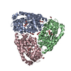
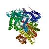
- PDB-4e3e: CRYSTAL STRUCTURE OF putative MaoC domain protein dehydratase fro... -
+
Open data
ID or keywords:
Loading...
-
Basic information
Entry
Database: PDB / ID: 4e3e
Title
CRYSTAL STRUCTURE OF putative MaoC domain protein dehydratase from Chloroflexus aurantiacus J-10-fl
Components
MaoC domain protein dehydratase
Keywords
OXIDOREDUCTASE / STRUCTURAL GENOMICS / PROTEIN STRUCTURE INITIATIVE / protein dehydratase / NYSGRC / PSI-Biology / New York Structural Genomics Research Consortium
Function / homology
Function and homology information
2-methylfumaryl-CoA hydratase / carbon fixation by 3-hydroxypropionate cycle / oxo-acid-lyase activity / glyoxylate catabolic process Similarity search - Function
Protocol: SINGLE WAVELENGTH / Scattering type: x-ray
Radiation wavelength
Wavelength: 0.9791 Å / Relative weight: 1
Reflection
Redundancy: 4.2 % / Av σ(I) over netI: 27.61 / Number: 742528 / Rmerge(I) obs: 0.069 / Χ2: 1.53 / D res high: 1.9 Å / D res low: 50 Å / Num. obs: 178479 / % possible obs: 99.8
Diffraction reflection shell
Highest resolution (Å)
Lowest resolution (Å)
% possible obs (%)
ID
Rmerge(I) obs
Chi squared
Redundancy
5.16
50
98.8
1
0.031
2.046
4.3
4.09
5.16
98.6
1
0.038
2.479
4.1
3.58
4.09
99.8
1
0.052
2.869
4.2
3.25
3.58
99.9
1
0.06
2.715
4.2
3.02
3.25
100
1
0.07
2.159
4.3
2.84
3.02
100
1
0.077
1.682
4.3
2.7
2.84
100
1
0.092
1.519
4.3
2.58
2.7
100
1
0.108
1.417
4.3
2.48
2.58
100
1
0.135
1.332
4.3
2.39
2.48
100
1
0.162
1.266
4.3
2.32
2.39
100
1
0.201
1.225
4.2
2.25
2.32
100
1
0.231
1.241
4.2
2.19
2.25
100
1
0.261
1.187
4.2
2.14
2.19
100
1
0.3
1.178
4.2
2.09
2.14
100
1
0.364
1.123
4.1
2.05
2.09
100
1
0.452
1.048
4.1
2.01
2.05
100
1
0.535
1.012
4.1
1.97
2.01
100
1
0.647
0.951
4
1.93
1.97
99.9
1
0.713
0.932
3.8
1.9
1.93
98.2
1
0.811
0.922
3.5
Reflection
Resolution: 1.9→50 Å / Num. obs: 178479 / % possible obs: 99.8 % / Redundancy: 4.2 % / Rmerge(I) obs: 0.069 / Χ2: 1.53 / Net I/σ(I): 10.3
Reflection shell
Resolution (Å)
Redundancy (%)
Rmerge(I) obs
Num. unique all
Χ2
Diffraction-ID
% possible all
1.9-1.93
3.5
0.811
8870
0.922
1
98.2
1.93-1.97
3.8
0.713
8850
0.932
1
99.9
1.97-2.01
4
0.647
8904
0.951
1
100
2.01-2.05
4.1
0.535
8996
1.012
1
100
2.05-2.09
4.1
0.452
8956
1.048
1
100
2.09-2.14
4.1
0.364
8962
1.123
1
100
2.14-2.19
4.2
0.3
8845
1.178
1
100
2.19-2.25
4.2
0.261
9044
1.187
1
100
2.25-2.32
4.2
0.231
8902
1.241
1
100
2.32-2.39
4.2
0.201
8926
1.225
1
100
2.39-2.48
4.3
0.162
8965
1.266
1
100
2.48-2.58
4.3
0.135
8935
1.332
1
100
2.58-2.7
4.3
0.108
8914
1.417
1
100
2.7-2.84
4.3
0.092
8976
1.519
1
100
2.84-3.02
4.3
0.077
8971
1.682
1
100
3.02-3.25
4.3
0.07
8909
2.159
1
100
3.25-3.58
4.2
0.06
8947
2.715
1
99.9
3.58-4.09
4.2
0.052
8928
2.869
1
99.8
4.09-5.16
4.1
0.038
8841
2.479
1
98.6
5.16-50
4.3
0.031
8838
2.046
1
98.8
-
Phasing
Phasing
Method: SAD
-
Processing
Software
Name
Version
Classification
NB
SCALEPACK
datascaling
RESOLVE
phasing
REFMAC
refinement
PDB_EXTRACT
3.1
dataextraction
CBASS
datacollection
HKL-3000
datareduction
PHENIX
phasing
Refinement
Method to determine structure: SAD / Resolution: 1.9→19.41 Å / Cor.coef. Fo:Fc: 0.981 / Cor.coef. Fo:Fc free: 0.967 / WRfactor Rfree: 0.183 / WRfactor Rwork: 0.1387 / Occupancy max: 1 / Occupancy min: 0.33 / FOM work R set: 0.8358 / SU B: 6.284 / SU ML: 0.078 / SU R Cruickshank DPI: 0.1194 / SU Rfree: 0.0967 / Cross valid method: THROUGHOUT / σ(F): 0 / ESU R: 0.119 / ESU R Free: 0.097 / Stereochemistry target values: MAXIMUM LIKELIHOOD Details: HYDROGENS HAVE BEEN USED IF PRESENT IN THE INPUT U VALUES : RESIDUAL ONLY
Rfactor
Num. reflection
% reflection
Selection details
Rfree
0.1883
4475
5 %
RANDOM
Rwork
0.1406
-
-
-
obs
0.143
89193
99.79 %
-
Solvent computation
Ion probe radii: 0.8 Å / Shrinkage radii: 0.8 Å / VDW probe radii: 1.2 Å / Solvent model: MASK
In the structure databanks used in Yorodumi, some data are registered as the other names, "COVID-19 virus" and "2019-nCoV". Here are the details of the virus and the list of structure data.
Jan 31, 2019. EMDB accession codes are about to change! (news from PDBe EMDB page)
EMDB accession codes are about to change! (news from PDBe EMDB page)
The allocation of 4 digits for EMDB accession codes will soon come to an end. Whilst these codes will remain in use, new EMDB accession codes will include an additional digit and will expand incrementally as the available range of codes is exhausted. The current 4-digit format prefixed with “EMD-” (i.e. EMD-XXXX) will advance to a 5-digit format (i.e. EMD-XXXXX), and so on. It is currently estimated that the 4-digit codes will be depleted around Spring 2019, at which point the 5-digit format will come into force.
The EM Navigator/Yorodumi systems omit the EMD- prefix.
Related info.:Q: What is EMD? / ID/Accession-code notation in Yorodumi/EM Navigator
Yorodumi is a browser for structure data from EMDB, PDB, SASBDB, etc.
This page is also the successor to EM Navigator detail page, and also detail information page/front-end page for Omokage search.
The word "yorodu" (or yorozu) is an old Japanese word meaning "ten thousand". "mi" (miru) is to see.
Related info.:EMDB / PDB / SASBDB / Comparison of 3 databanks / Yorodumi Search / Aug 31, 2016. New EM Navigator & Yorodumi / Yorodumi Papers / Jmol/JSmol / Function and homology information / Changes in new EM Navigator and Yorodumi
 Movie
Movie Controller

 Yorodumi
 Open data
 Basic information
 Components
 Keywords
 Function and homology information
 Chloroflexus aurantiacus (bacteria)
 X-RAY DIFFRACTION /
  Authors
 Citation
 Structure visualization
 Downloads & links
 Other downloads
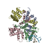


 PDBj
PDBj
 Assembly

 Mass: 96.063 Da / Num. of mol.: 2 / Source method: obtained synthetically / Formula: SO4
 Mass: 18.015 Da / Num. of mol.: 480 / Source method: isolated from a natural source / Formula: H2O
 Sample preparation
 / Beamline: X29A / Wavelength: 0.9791 Å
 Processing