 Movie
Movie Controller

[English] 日本語
 Yorodumi
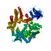
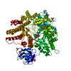
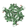
- PDB-3b2e: Crystal structure of S. cerevisiae Get3 in the open conformation ... -
+ Open data

- Basic information
| Entry | Database: PDB / ID: 3b2e | ||||||
|---|---|---|---|---|---|---|---|
| Title | Crystal structure of S. cerevisiae Get3 in the open conformation in complex with Get1 cytosolic domain | ||||||
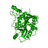
 Components Components |
| ||||||
 Keywords Keywords | HYDROLASE/TRANSPORT PROTEIN / Protein-protein interaction / receptor complex / HYDROLASE / TRANSPORT PROTEIN / ADP binding / Coild-coil / HYDROLASE-TRANSPORT PROTEIN complex | ||||||
| Function / homology |  Function and homology information Function and homology informationestablishment of protein localization to endoplasmic reticulum membrane / GET complex / : / tail-anchored membrane protein insertion into ER membrane / Hydrolases; Acting on acid anhydrides / protein insertion into ER membrane / post-translational protein targeting to endoplasmic reticulum membrane / retrograde vesicle-mediated transport, Golgi to endoplasmic reticulum / response to arsenic-containing substance / response to metal ion ...establishment of protein localization to endoplasmic reticulum membrane / GET complex / : / tail-anchored membrane protein insertion into ER membrane / Hydrolases; Acting on acid anhydrides / protein insertion into ER membrane / post-translational protein targeting to endoplasmic reticulum membrane / retrograde vesicle-mediated transport, Golgi to endoplasmic reticulum / response to arsenic-containing substance / response to metal ion / transmembrane protein transporter activity / mitophagy / protein-membrane adaptor activity / protein folding chaperone / guanyl-nucleotide exchange factor activity / mitochondrial membrane / : / response to heat / cellular response to oxidative stress / Golgi membrane / endoplasmic reticulum membrane / Golgi apparatus / endoplasmic reticulum / ATP hydrolysis activity / mitochondrion / ATP binding / metal ion binding / identical protein binding / cytosol Similarity search - Function | ||||||
| Biological species |  | ||||||
| Method |  X-RAY DIFFRACTION / SYNCHROTRON / MOLECULAR REPLACEMENT / Resolution: 3 Å X-RAY DIFFRACTION / SYNCHROTRON / MOLECULAR REPLACEMENT / Resolution: 3 Å | ||||||
 Authors Authors | Kubota, K. / Yamagata, A. / Fukai, S. | ||||||
 Citation Citation | Journal: J.Mol.Biol. / Year: 2012 Title: Get1 stabilizes an open dimer conformation of get3 ATPase by binding two distinct interfaces Authors: Kubota, K. / Yamagata, A. / Sato, Y. / Goto-Ito, S. / Fukai, S. | ||||||
| History |
|
- Structure visualization
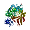
| Structure viewer | Molecule: MolmilJmol/JSmol |
|---|
- Downloads & links
-Download
| PDBx/mmCIF format | 3b2e.cif.gz | 309.9 KB | Display | PDBx/mmCIF format |
|---|---|---|---|---|
| PDB format | pdb3b2e.ent.gz | 250.3 KB | Display | PDB format |
| PDBx/mmJSON format | 3b2e.json.gz | Tree view | PDBx/mmJSON format | |
| Others |  Other downloads Other downloads |
-Validation report
| Arichive directory | https://data.pdbj.org/pub/pdb/validation_reports/b2/3b2eftp://data.pdbj.org/pub/pdb/validation_reports/b2/3b2e | HTTPS FTP |
|---|
-Related structure data
| Related structure data |  3vlcC  3a36S C: citing same article ( S: Starting model for refinement |
|---|---|
| Similar structure data |


-Links
 PDBj
PDBj- Assembly
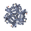
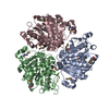
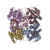
| Deposited unit | 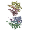
| ||||||||
|---|---|---|---|---|---|---|---|---|---|
| 1 | 
| ||||||||
| 2 | 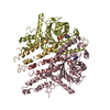
| ||||||||
| Unit cell |
|
-Components
| #1: Protein | Mass: 40522.766 Da / Num. of mol.: 4 Source method: isolated from a genetically manipulated source Source: (gene. exp.) Strain: NRRL Y-53 / Plasmid: PETDuet / Production host:  #2: Protein | Mass: 10064.349 Da / Num. of mol.: 4 / Fragment: Get1 cytosolic domain, UNP residues 20-103 Source method: isolated from a genetically manipulated source Source: (gene. exp.) Strain: ATCC 204508 / S288c / Gene: GET1 / Plasmid: PETDuet / Production host: #3: Chemical | ChemComp-ADP /   Mass: 427.201 Da / Num. of mol.: 4 / Source method: obtained synthetically / Formula: C10H15N5O10P2 / Comment: ADP, energy-carrying molecule*YM Mass: 427.201 Da / Num. of mol.: 4 / Source method: obtained synthetically / Formula: C10H15N5O10P2 / Comment: ADP, energy-carrying molecule*YMSequence details | A SEQUENCE DATABASE REFERENCE FOR CHAIN A, B, C, D WHICH DERIVES FROM STRAIN NRRL Y-53 DOES NOT ...A SEQUENCE DATABASE REFERENCE FOR CHAIN A, B, C, D WHICH DERIVES FROM STRAIN NRRL Y-53 DOES NOT CURRENTLY EXIST. | |
|---|
-Experimental details
-Experiment
| Experiment | Method: X-RAY DIFFRACTION / Number of used crystals: 1 |
|---|
- Sample preparation
| Crystal | Density Matthews: 2.9 Å3/Da / Density % sol: 57.53 % |
|---|---|
| Crystal grow | Temperature: 293 K / Method: vapor diffusion, sitting drop Details: 13.5% PEG3350, 0.18 M tri-sodium citrate, 9% MPD, VAPOR DIFFUSION, SITTING DROP, temperature 293K |
-Data collection
| Diffraction | Mean temperature: 100 K |
|---|---|
| Diffraction source | Source: SYNCHROTRON / Site: SPring-8  / Beamline: BL41XU / Wavelength: 1 Å / Beamline: BL41XU / Wavelength: 1 Å |
| Detector | Type: RAYONIX MX225HE / Detector: CCD / Date: May 21, 2011 |
| Radiation | Protocol: SINGLE WAVELENGTH / Monochromatic (M) / Laue (L): M / Scattering type: x-ray |
| Radiation wavelength | Wavelength: 1 Å / Relative weight: 1 |
| Reflection | Resolution: 3→50 Å / Num. obs: 44560 / % possible obs: 95 % / Observed criterion σ(F): 0 / Observed criterion σ(I): 0 / Redundancy: 4.5 % / Rmerge(I) obs: 0.096 / Net I/σ(I): 19.1 |
| Reflection shell | Resolution: 3→3.05 Å / Rmerge(I) obs: 0.469 / Mean I/σ(I) obs: 2 / % possible all: 87.2 |
- Processing
| Software |
| ||||||||||||||||||||
|---|---|---|---|---|---|---|---|---|---|---|---|---|---|---|---|---|---|---|---|---|---|
| Refinement | Method to determine structure: MOLECULAR REPLACEMENT Starting model: 3A36 Resolution: 3→50 Å / σ(F): 0 / Stereochemistry target values: MAXIMUM LIKELIHOOD
| ||||||||||||||||||||
| Refinement step | Cycle: LAST / Resolution: 3→50 Å
| ||||||||||||||||||||
| Refine LS restraints |
|
