 Movie
Movie Controller


+ Open data

- Basic information
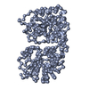
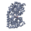
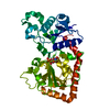
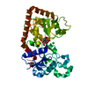
| Entry | Database: PDB / ID: 1jix | ||||||
|---|---|---|---|---|---|---|---|
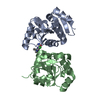
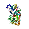
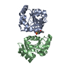
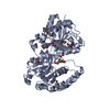
| Title | T4 Phage BGT in Complex with Ca2+ | ||||||
 Components Components | DNA BETA-GLUCOSYLTRANSFERASE | ||||||
 Keywords Keywords | TRANSFERASE / Glycosyltransferase | ||||||
| Function / homology |  Function and homology information Function and homology informationDNA beta-glucosyltransferase / DNA beta-glucosyltransferase activity / symbiont-mediated evasion of host restriction-modification system / DNA modification / symbiont-mediated suppression of host innate immune response Similarity search - Function | ||||||
| Biological species |  Enterobacteria phage T4 (virus) Enterobacteria phage T4 (virus) | ||||||
| Method |  X-RAY DIFFRACTION / SYNCHROTRON / MOLECULAR REPLACEMENT / Resolution: 1.65 Å X-RAY DIFFRACTION / SYNCHROTRON / MOLECULAR REPLACEMENT / Resolution: 1.65 Å | ||||||
 Authors Authors | Morera, S. / Lariviere, L. / Kurzeck, J. / Aschke-Sonnenborn, U. / Freemont, P.S. / Janin, J. / Ruger, W. | ||||||
 Citation Citation | Journal: J.Mol.Biol. / Year: 2001 Title: High resolution crystal structures of T4 phage beta-glucosyltransferase: induced fit and effect of substrate and metal binding. Authors: Morera, S. / Lariviere, L. / Kurzeck, J. / Aschke-Sonnenborn, U. / Freemont, P.S. / Janin, J. / Ruger, W. #1: Journal: J.Mol.Biol. / Year: 1999Title: T4 phage beta-glucosyltransferase : substrate binding and proposed catalytic mechanism Authors: Morera, S. / Imberty, A. / Aschke-Sonnenborn, U. / Ruger, W. / Freemont, P.S. | ||||||
| History |
|
- Structure visualization
| Structure viewer | Molecule: MolmilJmol/JSmol |
|---|
- Downloads & links
-Download
| PDBx/mmCIF format | 1jix.cif.gz | 96.1 KB | Display | PDBx/mmCIF format |
|---|---|---|---|---|
| PDB format | pdb1jix.ent.gz | 70.8 KB | Display | PDB format |
| PDBx/mmJSON format | 1jix.json.gz | Tree view | PDBx/mmJSON format | |
| Others |  Other downloads Other downloads |
-Validation report
| Arichive directory | https://data.pdbj.org/pub/pdb/validation_reports/ji/1jixftp://data.pdbj.org/pub/pdb/validation_reports/ji/1jix | HTTPS FTP |
|---|
-Related structure data
| Related structure data | 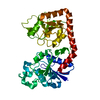 1jejC  1jg6C 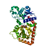 1jg7C 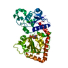 1jiuC  1jivC 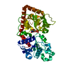 1qkjS S: Starting model for refinement C: citing same article ( |
|---|---|
| Similar structure data |


-Links
 PDBj
PDBj


- Assembly
| Deposited unit | 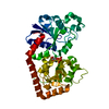
| ||||||||
|---|---|---|---|---|---|---|---|---|---|
| 1 |
| ||||||||
| Unit cell |
|
-Components
| #1: Protein | Mass: 40719.879 Da / Num. of mol.: 1 Source method: isolated from a genetically manipulated source Source: (gene. exp.) Enterobacteria phage T4 (virus) / Genus: T4-like viruses / Species: Enterobacteria phage T4 sensu lato / Production host:  |
|---|---|
| #2: Chemical | ChemComp-CA /   Mass: 40.078 Da / Num. of mol.: 1 / Source method: obtained synthetically / Formula: Ca Mass: 40.078 Da / Num. of mol.: 1 / Source method: obtained synthetically / Formula: Ca |
| #3: Chemical | ChemComp-UDP /   Type: RNA linking / Mass: 404.161 Da / Num. of mol.: 1 / Source method: obtained synthetically / Formula: C9H14N2O12P2 / Comment: UDP*YM Type: RNA linking / Mass: 404.161 Da / Num. of mol.: 1 / Source method: obtained synthetically / Formula: C9H14N2O12P2 / Comment: UDP*YM |
| #4: Water | ChemComp-HOH /  Mass: 18.015 Da / Num. of mol.: 434 / Source method: isolated from a natural source / Formula: H2O Mass: 18.015 Da / Num. of mol.: 434 / Source method: isolated from a natural source / Formula: H2O |
-Experimental details
-Experiment
| Experiment | Method: X-RAY DIFFRACTION / Number of used crystals: 1 |
|---|
- Sample preparation
| Crystal | Density Matthews: 2.61 Å3/Da / Density % sol: 52.82 % | ||||||||||||||||||||||||||||||
|---|---|---|---|---|---|---|---|---|---|---|---|---|---|---|---|---|---|---|---|---|---|---|---|---|---|---|---|---|---|---|---|
| Crystal grow | Temperature: 291 K / Method: vapor diffusion, hanging drop / pH: 7.5 Details: PEG 10000, pH 7.5, VAPOR DIFFUSION, HANGING DROP, temperature 291K | ||||||||||||||||||||||||||||||
| Crystal grow | *PLUS | ||||||||||||||||||||||||||||||
| Components of the solutions | *PLUS
|
-Data collection
| Diffraction | Mean temperature: 100 K |
|---|---|
| Diffraction source | Source: SYNCHROTRON / Site: ESRF  / Beamline: ID14-1 / Wavelength: 0.934 Å / Beamline: ID14-1 / Wavelength: 0.934 Å |
| Detector | Type: MARRESEARCH / Detector: CCD / Date: Sep 7, 2000 |
| Radiation | Protocol: SINGLE WAVELENGTH / Monochromatic (M) / Laue (L): M / Scattering type: x-ray |
| Radiation wavelength | Wavelength: 0.934 Å / Relative weight: 1 |
| Reflection | Resolution: 1.65→20 Å / Num. all: 50720 / Num. obs: 49706 / % possible obs: 98.8 % / Observed criterion σ(F): 2 / Observed criterion σ(I): 1 / Redundancy: 6.1 % / Biso Wilson estimate: 20 Å2 / Rmerge(I) obs: 0.04 / Net I/σ(I): 10 |
| Reflection shell | Resolution: 1.65→1.71 Å / Rmerge(I) obs: 0.149 / Mean I/σ(I) obs: 6.5 / Num. unique all: 4908 / % possible all: 97.1 |
| Reflection | *PLUS Lowest resolution: 20 Å / Num. measured all: 304335 / Rmerge(I) obs: 0.04 |
- Processing
| Software |
| |||||||||||||||||||||||||
|---|---|---|---|---|---|---|---|---|---|---|---|---|---|---|---|---|---|---|---|---|---|---|---|---|---|---|
| Refinement | Method to determine structure: MOLECULAR REPLACEMENT Starting model: PDB ENTRY 1QKJ Resolution: 1.65→20 Å / σ(F): 2 / σ(I): 0 / Stereochemistry target values: Engh & Huber
| |||||||||||||||||||||||||
| Refinement step | Cycle: LAST / Resolution: 1.65→20 Å
| |||||||||||||||||||||||||
| Refine LS restraints |
| |||||||||||||||||||||||||
| Software | *PLUS Name: CNS / Version: 1 / Classification: refinement | |||||||||||||||||||||||||
| Refinement | *PLUS Lowest resolution: 20 Å / σ(F): 2 / % reflection Rfree: 5 % / Rfactor obs: 0.2 / Rfactor Rwork: 0.2 | |||||||||||||||||||||||||
| Solvent computation | *PLUS | |||||||||||||||||||||||||
| Displacement parameters | *PLUS |
