 Movie
Movie Controller


+ Open data

- Basic information
| Entry | Database: PDB / ID: 1nzf | ||||||
|---|---|---|---|---|---|---|---|
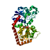
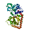
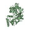
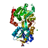
| Title | T4 phage BGT-D100A mutant in complex with UDP-glucose: Form II | ||||||
 Components Components | DNA beta-glycosyltransferase | ||||||
 Keywords Keywords | TRANSFERASE / glycosyltransferase / GT-B / UDP-glucose | ||||||
| Function / homology |  Function and homology information Function and homology informationDNA beta-glucosyltransferase / DNA beta-glucosyltransferase activity / symbiont-mediated evasion of host restriction-modification system / DNA modification / symbiont-mediated suppression of host innate immune response Similarity search - Function | ||||||
| Biological species |  Enterobacteria phage T4 (virus) Enterobacteria phage T4 (virus) | ||||||
| Method |  X-RAY DIFFRACTION / SYNCHROTRON / MOLECULAR REPLACEMENT / Resolution: 2.1 Å X-RAY DIFFRACTION / SYNCHROTRON / MOLECULAR REPLACEMENT / Resolution: 2.1 Å | ||||||
 Authors Authors | Lariviere, L. / Morera, S. | ||||||
 Citation Citation | Journal: J.Mol.Biol. / Year: 2003 Title: Crystal structures of the T4 phage beta-glucosyltransferase and the D100A mutant in complex with UDP-glucose: glucose binding and identification of the catalytic base for a direct displacement mechanism. Authors: Lariviere, L. / Gueguen-Chaignon, V. / Morera, S. | ||||||
| History |
|
- Structure visualization
| Structure viewer | Molecule: MolmilJmol/JSmol |
|---|
- Downloads & links
-Download
| PDBx/mmCIF format | 1nzf.cif.gz | 94 KB | Display | PDBx/mmCIF format |
|---|---|---|---|---|
| PDB format | pdb1nzf.ent.gz | 69.6 KB | Display | PDB format |
| PDBx/mmJSON format | 1nzf.json.gz | Tree view | PDBx/mmJSON format | |
| Others |  Other downloads Other downloads |
-Validation report
| Arichive directory | https://data.pdbj.org/pub/pdb/validation_reports/nz/1nzfftp://data.pdbj.org/pub/pdb/validation_reports/nz/1nzf | HTTPS FTP |
|---|
-Related structure data
| Related structure data | 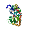 1j39SC  1nvkC 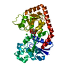 1nzdC S: Starting model for refinement C: citing same article ( |
|---|---|
| Similar structure data |


-Links
 PDBj
PDBj
- Assembly
| Deposited unit | 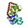
| ||||||||
|---|---|---|---|---|---|---|---|---|---|
| 1 |
| ||||||||
| Unit cell |
|
-Components
| #1: Protein | Mass: 40675.871 Da / Num. of mol.: 1 / Mutation: D100A Source method: isolated from a genetically manipulated source Source: (gene. exp.) Enterobacteria phage T4 (virus) / Genus: T4-like viruses / Species: Enterobacteria phage T4 sensu lato / Gene: BGT / Plasmid: pBSK / Species (production host): Escherichia coli / Production host:  | ||||||
|---|---|---|---|---|---|---|---|
| #2: Chemical |   Mass: 35.453 Da / Num. of mol.: 2 / Source method: obtained synthetically / Formula: Cl Mass: 35.453 Da / Num. of mol.: 2 / Source method: obtained synthetically / Formula: Cl#3: Chemical | ChemComp-UPG / |   Mass: 566.302 Da / Num. of mol.: 1 / Source method: obtained synthetically / Formula: C15H24N2O17P2 Mass: 566.302 Da / Num. of mol.: 1 / Source method: obtained synthetically / Formula: C15H24N2O17P2#4: Chemical |   Mass: 92.094 Da / Num. of mol.: 2 / Source method: obtained synthetically / Formula: C3H8O3 Mass: 92.094 Da / Num. of mol.: 2 / Source method: obtained synthetically / Formula: C3H8O3#5: Water | ChemComp-HOH / |  Mass: 18.015 Da / Num. of mol.: 337 / Source method: isolated from a natural source / Formula: H2O Mass: 18.015 Da / Num. of mol.: 337 / Source method: isolated from a natural source / Formula: H2O |
-Experimental details
-Experiment
| Experiment | Method: X-RAY DIFFRACTION / Number of used crystals: 1 |
|---|
- Sample preparation
| Crystal | Density Matthews: 2.02 Å3/Da / Density % sol: 38.65 % |
|---|---|
| Crystal grow | Temperature: 292 K / Method: vapor diffusion, hanging drop / pH: 7.5 Details: peg 4000, glycerol, pH 7.5, VAPOR DIFFUSION, HANGING DROP, temperature 292K |
-Data collection
| Diffraction | Mean temperature: 100 K |
|---|---|
| Diffraction source | Source: SYNCHROTRON / Site: ESRF  / Beamline: BM30A / Wavelength: 0.97 Å / Beamline: BM30A / Wavelength: 0.97 Å |
| Detector | Type: MARRESEARCH / Detector: CCD / Date: Feb 10, 2003 |
| Radiation | Monochromator: 0.95 / Protocol: SINGLE WAVELENGTH / Monochromatic (M) / Laue (L): M / Scattering type: x-ray |
| Radiation wavelength | Wavelength: 0.97 Å / Relative weight: 1 |
| Reflection | Resolution: 2.1→20 Å / Num. all: 22179 / Num. obs: 21948 / % possible obs: 99 % / Observed criterion σ(F): 0 / Observed criterion σ(I): 0 / Biso Wilson estimate: 28 Å2 / Rsym value: 0.05 / Net I/σ(I): 18.7 |
| Reflection shell | Resolution: 2.1→2.18 Å / Mean I/σ(I) obs: 5.5 / Num. unique all: 2146 / Rsym value: 0.3 / % possible all: 99 |
- Processing
| Software |
| ||||||||||||||||||||||||||||
|---|---|---|---|---|---|---|---|---|---|---|---|---|---|---|---|---|---|---|---|---|---|---|---|---|---|---|---|---|---|
| Refinement | Method to determine structure: MOLECULAR REPLACEMENT Starting model: PDB ENTRY 1J39 Resolution: 2.1→20 Å / Cross valid method: THROUGHOUT / σ(F): 0 / Stereochemistry target values: Engh & Huber
| ||||||||||||||||||||||||||||
| Displacement parameters |
| ||||||||||||||||||||||||||||
| Refinement step | Cycle: LAST / Resolution: 2.1→20 Å
| ||||||||||||||||||||||||||||
| Refine LS restraints |
| ||||||||||||||||||||||||||||
| Xplor file |
|
