 Movie
Movie Controller


+ Open data

- Basic information
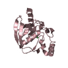
| Entry | Database: PDB / ID: 1j7j | ||||||
|---|---|---|---|---|---|---|---|
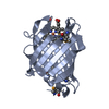
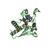
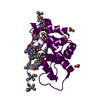
| Title | Crystal Structure of the HPRT from Salmonella typhimurium | ||||||
 Components Components | hypoxanthine phosphoribosyltransferase | ||||||
 Keywords Keywords | TRANSFERASE / GLYCOSYLTRANSFERASE / PHOSPHORIBOSYLTRANSFERASE / NUCLEOTIDE METABOLISM / PURINE SALVAGE | ||||||
| Function / homology |  Function and homology information Function and homology informationhypoxanthine phosphoribosyltransferase / guanine phosphoribosyltransferase activity / guanine salvage / hypoxanthine metabolic process / hypoxanthine phosphoribosyltransferase activity / GMP salvage / IMP salvage / purine ribonucleoside salvage / nucleotide binding / magnesium ion binding / cytosol Similarity search - Function | ||||||
| Biological species |  Salmonella typhimurium (bacteria) Salmonella typhimurium (bacteria) | ||||||
| Method |  X-RAY DIFFRACTION / SYNCHROTRON / MOLECULAR REPLACEMENT / Resolution: 2.3 Å X-RAY DIFFRACTION / SYNCHROTRON / MOLECULAR REPLACEMENT / Resolution: 2.3 Å | ||||||
 Authors Authors | Lee, C.C. / Focia, P.J. / Spraggon, G. / Eakin, A.E. | ||||||
 Citation Citation | Journal: To be Published Title: Crystal Structure of the HPRT from Salmonella Typhimurium at 2.3 A Resolution Authors: Lee, C.C. / Focia, P.J. / Spraggon, G. / Eakin, A.E. | ||||||
| History |
|
- Structure visualization
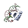
| Structure viewer | Molecule: MolmilJmol/JSmol |
|---|
- Downloads & links
-Download
| PDBx/mmCIF format | 1j7j.cif.gz | 79.5 KB | Display | PDBx/mmCIF format |
|---|---|---|---|---|
| PDB format | pdb1j7j.ent.gz | 60.2 KB | Display | PDB format |
| PDBx/mmJSON format | 1j7j.json.gz | Tree view | PDBx/mmJSON format | |
| Others |  Other downloads Other downloads |
-Validation report
| Arichive directory | https://data.pdbj.org/pub/pdb/validation_reports/j7/1j7jftp://data.pdbj.org/pub/pdb/validation_reports/j7/1j7j | HTTPS FTP |
|---|
-Related structure data
| Related structure data | 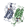 1tc1S S: Starting model for refinement |
|---|---|
| Similar structure data |


-Links
 PDBj
PDBj
- Assembly
| Deposited unit | 
| |||||||||
|---|---|---|---|---|---|---|---|---|---|---|
| 1 | 
| |||||||||
| 2 | 
| |||||||||
| 3 | 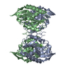
| |||||||||
| Unit cell |
| |||||||||
| Components on special symmetry positions |
|
-Components
| #1: Protein | Mass: 20094.191 Da / Num. of mol.: 2 Source method: isolated from a genetically manipulated source Source: (gene. exp.) Salmonella typhimurium (bacteria) / Gene: hpt / Plasmid: pBAce / Production host: References: UniProt: O33799, hypoxanthine phosphoribosyltransferase #2: Chemical |   Mass: 24.305 Da / Num. of mol.: 2 / Source method: obtained synthetically / Formula: Mg Mass: 24.305 Da / Num. of mol.: 2 / Source method: obtained synthetically / Formula: Mg#3: Water | ChemComp-HOH / |  Mass: 18.015 Da / Num. of mol.: 132 / Source method: isolated from a natural source / Formula: H2O Mass: 18.015 Da / Num. of mol.: 132 / Source method: isolated from a natural source / Formula: H2O |
|---|
-Experimental details
-Experiment
| Experiment | Method: X-RAY DIFFRACTION / Number of used crystals: 1 |
|---|
- Sample preparation
| Crystal | Density Matthews: 4.26 Å3/Da / Density % sol: 71.16 % |
|---|---|
| Crystal grow | pH: 7.5 / Details: 28% PEG 400, 200mM CaCl2, 100mM HEPES pH 7.5 |
-Data collection
| Diffraction | Mean temperature: 100 K |
|---|---|
| Diffraction source | Source: SYNCHROTRON / Site: APS  / Beamline: 5ID-B / Wavelength: 1 / Beamline: 5ID-B / Wavelength: 1 |
| Detector | Type: MAR 165 mm CCD / Detector: CCD / Date: Nov 14, 1998 |
| Radiation | Protocol: SINGLE WAVELENGTH / Monochromatic (M) / Laue (L): M / Scattering type: x-ray |
| Radiation wavelength | Wavelength: 1 Å / Relative weight: 1 |
| Reflection | Resolution: 2.3→40 Å / Num. obs: 31306 / % possible obs: 99.8 % / Observed criterion σ(I): -3 / Redundancy: 10.8 % / Rmerge(I) obs: 0.05 / Rsym value: 0.05 / Net I/σ(I): 46.4 |
| Reflection shell | Resolution: 2.3→2.35 Å / Redundancy: 11 % / Rmerge(I) obs: 0.301 / Mean I/σ(I) obs: 7.7 / Rsym value: 0.301 / % possible all: 100 |
- Processing
| Software |
| ||||||||||||||||||||||||||||||||||||||||||||||||||||||||||||
|---|---|---|---|---|---|---|---|---|---|---|---|---|---|---|---|---|---|---|---|---|---|---|---|---|---|---|---|---|---|---|---|---|---|---|---|---|---|---|---|---|---|---|---|---|---|---|---|---|---|---|---|---|---|---|---|---|---|---|---|---|---|
| Refinement | Method to determine structure: MOLECULAR REPLACEMENT Starting model: 1TC1 without ligands and waters Resolution: 2.3→500 Å
| ||||||||||||||||||||||||||||||||||||||||||||||||||||||||||||
| Refinement step | Cycle: LAST / Resolution: 2.3→500 Å
| ||||||||||||||||||||||||||||||||||||||||||||||||||||||||||||
| Refine LS restraints |
|
