 Movie
Movie Controller

[English] 日本語
 Yorodumi
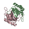
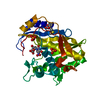
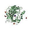
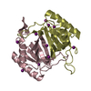
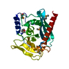
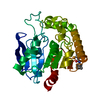
- PDB-1j7g: Structure of YihZ from Haemophilus influenzae (HI0670), a D-Tyr-t... -
+ Open data

- Basic information
| Entry | Database: PDB / ID: 1j7g | ||||||
|---|---|---|---|---|---|---|---|
| Title | Structure of YihZ from Haemophilus influenzae (HI0670), a D-Tyr-tRNA(Tyr) deacylase | ||||||
 Components Components | D-tyrosyl-tRNA(Tyr) deacylase | ||||||
 Keywords Keywords | HYDROLASE / D-Tyr-tRNA(Tyr) deacylase / structural genomics / hypothetical protein / Structure 2 Function Project / S2F | ||||||
| Function / homology |  Function and homology information Function and homology informationSer(Gly)-tRNA(Ala) hydrolase activity / D-tyrosyl-tRNA(Tyr) deacylase activity / Gly-tRNA(Ala) deacylase activity / D-aminoacyl-tRNA deacylase / tRNA metabolic process / D-amino acid catabolic process / tRNA binding / cytoplasm Similarity search - Function | ||||||
| Biological species |  Haemophilus influenzae Rd KW20 (bacteria) Haemophilus influenzae Rd KW20 (bacteria) | ||||||
| Method |  X-RAY DIFFRACTION / SYNCHROTRON / SAS / Resolution: 1.64 Å X-RAY DIFFRACTION / SYNCHROTRON / SAS / Resolution: 1.64 Å | ||||||
 Authors Authors | Lim, K. / Herzberg, O. / Structure 2 Function Project (S2F) | ||||||
 Citation Citation | Journal: J.Biol.Chem. / Year: 2003 Title: A Catalytic Mechanism for D-Tyr-tRNATyr Deacylase Based on the Crystal Structure of Hemophilus influenzae HI0670 Authors: Lim, K. / Tempczyk, A. / Bonander, N. / Toedt, J. / Howard, A. / Einsenstein, E. / Herzberg, O. #1: Journal: J.Biol.Chem. / Year: 1999Title: Functional Characterization of the D-Tyr-tRNA(Tyr) Deacylase from Escherichia coli Authors: Soutourina, J. / Plateau, P. / Delort, F. / Peirotes, A. / Blanquet, S. | ||||||
| History |
|
- Structure visualization
| Structure viewer | Molecule: MolmilJmol/JSmol |
|---|
- Downloads & links
-Download
| PDBx/mmCIF format | 1j7g.cif.gz | 43.9 KB | Display | PDBx/mmCIF format |
|---|---|---|---|---|
| PDB format | pdb1j7g.ent.gz | 30.9 KB | Display | PDB format |
| PDBx/mmJSON format | 1j7g.json.gz | Tree view | PDBx/mmJSON format | |
| Others |  Other downloads Other downloads |
-Validation report
| Arichive directory | https://data.pdbj.org/pub/pdb/validation_reports/j7/1j7gftp://data.pdbj.org/pub/pdb/validation_reports/j7/1j7g | HTTPS FTP |
|---|
-Related structure data
| Similar structure data | |
|---|---|
| Other databases |


-Links
 PDBj
PDBj- Assembly
| Deposited unit | 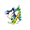
| ||||||||
|---|---|---|---|---|---|---|---|---|---|
| 1 | 
| ||||||||
| Unit cell |
|
-Components
| #1: Protein | Mass: 15879.174 Da / Num. of mol.: 1 Source method: isolated from a genetically manipulated source Source: (gene. exp.) Haemophilus influenzae Rd KW20 (bacteria)Species: Haemophilus influenzae / Strain: Kw20 / Gene: HI0670 / Plasmid: pET15b-HI0670 / Species (production host): Escherichia coli / Production host: References: UniProt: P44814, Hydrolases; Acting on ester bonds |
|---|---|
| #2: Water | ChemComp-HOH /  Mass: 18.015 Da / Num. of mol.: 207 / Source method: isolated from a natural source / Formula: H2O Mass: 18.015 Da / Num. of mol.: 207 / Source method: isolated from a natural source / Formula: H2O |
-Experimental details
-Experiment
| Experiment | Method: X-RAY DIFFRACTION / Number of used crystals: 1 |
|---|
- Sample preparation
| Crystal | Density Matthews: 3.12 Å3/Da / Density % sol: 60.55 % | ||||||||||||||||||||||||||||||
|---|---|---|---|---|---|---|---|---|---|---|---|---|---|---|---|---|---|---|---|---|---|---|---|---|---|---|---|---|---|---|---|
| Crystal grow | Temperature: 277 K Method: crystal formed during dialysis of protein (2.8mg/ml) pH: 7.5 Details: 50 mM Tris, 0.1 mM DTT, 0.1 mM EDTA, pH 7.5, crystal formed during dialysis of protein (2.8mg/ml), temperature 277K | ||||||||||||||||||||||||||||||
| Crystal grow | *PLUS Temperature: 4 ℃ / Method: vapor diffusion | ||||||||||||||||||||||||||||||
| Components of the solutions | *PLUS
|
-Data collection
| Diffraction | Mean temperature: 100 K |
|---|---|
| Diffraction source | Source: SYNCHROTRON / Site: APS  / Beamline: 17-ID / Wavelength: 1 Å / Beamline: 17-ID / Wavelength: 1 Å |
| Detector | Type: MARRESEARCH / Detector: CCD / Date: Jan 1, 2000 |
| Radiation | Protocol: SINGLE WAVELENGTH / Monochromatic (M) / Laue (L): M / Scattering type: x-ray |
| Radiation wavelength | Wavelength: 1 Å / Relative weight: 1 |
| Reflection | Resolution: 1.64→50 Å / Num. all: 25510 / Num. obs: 25510 / % possible obs: 99.8 % / Observed criterion σ(F): 0 / Observed criterion σ(I): 0 / Redundancy: 13 % / Biso Wilson estimate: 19 Å2 / Rmerge(I) obs: 0.037 / Net I/σ(I): 12.8 |
| Reflection shell | Resolution: 1.64→1.71 Å / Redundancy: 9.4 % / Rmerge(I) obs: 0.135 / % possible all: 100 |
| Reflection | *PLUS Lowest resolution: 50 Å / Num. measured all: 352028 |
| Reflection shell | *PLUS % possible obs: 100 % |
- Processing
| Software |
| ||||||||||||||||||||
|---|---|---|---|---|---|---|---|---|---|---|---|---|---|---|---|---|---|---|---|---|---|
| Refinement | Method to determine structure: SAS / Resolution: 1.64→20 Å / σ(F): 2 / Stereochemistry target values: Engh & Huber
| ||||||||||||||||||||
| Displacement parameters | Biso mean: 27 Å2 | ||||||||||||||||||||
| Refinement step | Cycle: LAST / Resolution: 1.64→20 Å
| ||||||||||||||||||||
| Refine LS restraints |
| ||||||||||||||||||||
| LS refinement shell | Resolution: 1.64→1.71 Å / Rfactor Rfree: 0.276 / Rfactor Rwork: 0.238 | ||||||||||||||||||||
| Refinement | *PLUS Rfactor Rfree: 0.22 | ||||||||||||||||||||
| Solvent computation | *PLUS | ||||||||||||||||||||
| Displacement parameters | *PLUS | ||||||||||||||||||||
| LS refinement shell | *PLUS Rfactor Rfree: 0.277 / Rfactor Rwork: 0.237 |
