 Movie
Movie Controller

[English] 日本語
 Yorodumi
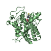
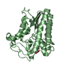
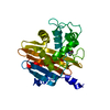
- PDB-6uqw: Crystal structure of ChoE in complex with acetate and thiocholine -
+ Open data

- Basic information
| Entry | Database: PDB / ID: 6uqw | ||||||
|---|---|---|---|---|---|---|---|
| Title | Crystal structure of ChoE in complex with acetate and thiocholine | ||||||
 Components Components | ChoE | ||||||
 Keywords Keywords | HYDROLASE / esterase / acetylcholine | ||||||
| Function / homology |  Function and homology information Function and homology information | ||||||
| Biological species |   Pseudomonas aeruginosa (bacteria) Pseudomonas aeruginosa (bacteria) | ||||||
| Method |  X-RAY DIFFRACTION / SYNCHROTRON / MOLECULAR REPLACEMENT / Resolution: 1.65 Å X-RAY DIFFRACTION / SYNCHROTRON / MOLECULAR REPLACEMENT / Resolution: 1.65 Å | ||||||
 Authors Authors | Pham, V.D. / Shi, R. | ||||||
| Funding support |  Canada, 1items Canada, 1items
| ||||||
 Citation Citation | Journal: J.Biol.Chem. / Year: 2020 Title: Structural insights into the putative bacterial acetylcholinesterase ChoE and its substrate inhibition mechanism. Authors: Pham, V.D. / To, T.A. / Gagne-Thivierge, C. / Couture, M. / Lague, P. / Yao, D. / Picard, M.E. / Lortie, L.A. / Attere, S.A. / Zhu, X. / Levesque, R.C. / Charette, S.J. / Shi, R. | ||||||
| History |
|
- Structure visualization
| Structure viewer | Molecule: MolmilJmol/JSmol |
|---|
- Downloads & links
-Download
| PDBx/mmCIF format | 6uqw.cif.gz | 138.8 KB | Display | PDBx/mmCIF format |
|---|---|---|---|---|
| PDB format | pdb6uqw.ent.gz | 105.8 KB | Display | PDB format |
| PDBx/mmJSON format | 6uqw.json.gz | Tree view | PDBx/mmJSON format | |
| Others |  Other downloads Other downloads |
-Validation report
| Arichive directory | https://data.pdbj.org/pub/pdb/validation_reports/uq/6uqwftp://data.pdbj.org/pub/pdb/validation_reports/uq/6uqw | HTTPS FTP |
|---|
-Related structure data
| Related structure data |  6uqvSC 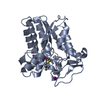 6uqxC 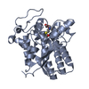 6uqyC 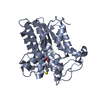 6uqzC 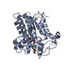 6ur0C 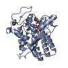 6ur1C S: Starting model for refinement C: citing same article ( |
|---|---|
| Similar structure data |


-Links
 PDBj
PDBj


- Assembly
| Deposited unit | 
| ||||||||
|---|---|---|---|---|---|---|---|---|---|
| 1 | 
| ||||||||
| 2 | 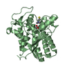
| ||||||||
| Unit cell |
|
-Components
| #1: Protein | Mass: 31543.293 Da / Num. of mol.: 2 / Fragment: UNP residues 21-307 Source method: isolated from a genetically manipulated source Source: (gene. exp.) Pseudomonas aeruginosa (strain ATCC 15692 / DSM 22644 / CIP 104116 / JCM 14847 / LMG 12228 / 1C / PRS 101 / PAO1) (bacteria)Strain: ATCC 15692 / DSM 22644 / CIP 104116 / JCM 14847 / LMG 12228 / 1C / PRS 101 / PAO1 Gene: choE, PA4921 / Plasmid: pET28a / Production host: References: UniProt: Q9HUP2, Hydrolases; Acting on ester bonds #2: Chemical |   Mass: 59.044 Da / Num. of mol.: 2 / Source method: obtained synthetically / Formula: C2H3O2 / Feature type: SUBJECT OF INVESTIGATION Mass: 59.044 Da / Num. of mol.: 2 / Source method: obtained synthetically / Formula: C2H3O2 / Feature type: SUBJECT OF INVESTIGATION#3: Chemical |   Mass: 120.236 Da / Num. of mol.: 3 / Source method: obtained synthetically / Formula: C5H14NS / Feature type: SUBJECT OF INVESTIGATION Mass: 120.236 Da / Num. of mol.: 3 / Source method: obtained synthetically / Formula: C5H14NS / Feature type: SUBJECT OF INVESTIGATION#4: Chemical | ChemComp-GOL / |   Mass: 92.094 Da / Num. of mol.: 1 / Source method: obtained synthetically / Formula: C3H8O3 Mass: 92.094 Da / Num. of mol.: 1 / Source method: obtained synthetically / Formula: C3H8O3#5: Water | ChemComp-HOH / |  Mass: 18.015 Da / Num. of mol.: 593 / Source method: isolated from a natural source / Formula: H2O Mass: 18.015 Da / Num. of mol.: 593 / Source method: isolated from a natural source / Formula: H2OHas ligand of interest | Y | Has protein modification | Y | |
|---|
-Experimental details
-Experiment
| Experiment | Method: X-RAY DIFFRACTION / Number of used crystals: 1 |
|---|
- Sample preparation
| Crystal | Density Matthews: 2.38 Å3/Da / Density % sol: 48.32 % / Mosaicity: 0.67 ° |
|---|---|
| Crystal grow | Temperature: 277 K / Method: microbatch / pH: 6.5 / Details: 15% PEG20000, 0.1 M MES, pH 6.5 |
-Data collection
| Diffraction | Mean temperature: 100 K / Serial crystal experiment: N | |||||||||||||||||||||||||||||||||||||||||||||||||||||||||||||||||||||||||||||||||||||||||||||||||||||||||||||||||||||||||
|---|---|---|---|---|---|---|---|---|---|---|---|---|---|---|---|---|---|---|---|---|---|---|---|---|---|---|---|---|---|---|---|---|---|---|---|---|---|---|---|---|---|---|---|---|---|---|---|---|---|---|---|---|---|---|---|---|---|---|---|---|---|---|---|---|---|---|---|---|---|---|---|---|---|---|---|---|---|---|---|---|---|---|---|---|---|---|---|---|---|---|---|---|---|---|---|---|---|---|---|---|---|---|---|---|---|---|---|---|---|---|---|---|---|---|---|---|---|---|---|---|---|---|
| Diffraction source | Source: SYNCHROTRON / Site: APS  / Beamline: 31-ID / Wavelength: 0.97931 Å / Beamline: 31-ID / Wavelength: 0.97931 Å | |||||||||||||||||||||||||||||||||||||||||||||||||||||||||||||||||||||||||||||||||||||||||||||||||||||||||||||||||||||||||
| Detector | Type: DECTRIS PILATUS3 S 6M / Detector: PIXEL / Date: Jun 19, 2019 | |||||||||||||||||||||||||||||||||||||||||||||||||||||||||||||||||||||||||||||||||||||||||||||||||||||||||||||||||||||||||
| Radiation | Monochromator: diamond(111) / Protocol: SINGLE WAVELENGTH / Monochromatic (M) / Laue (L): M / Scattering type: x-ray | |||||||||||||||||||||||||||||||||||||||||||||||||||||||||||||||||||||||||||||||||||||||||||||||||||||||||||||||||||||||||
| Radiation wavelength | Wavelength: 0.97931 Å / Relative weight: 1 | |||||||||||||||||||||||||||||||||||||||||||||||||||||||||||||||||||||||||||||||||||||||||||||||||||||||||||||||||||||||||
| Reflection | Resolution: 1.65→80.173 Å / Num. all: 68456 / Num. obs: 68456 / % possible obs: 96.5 % / Redundancy: 3.2 % / Rpim(I) all: 0.067 / Rrim(I) all: 0.123 / Rsym value: 0.103 / Net I/av σ(I): 4.9 / Net I/σ(I): 7.6 / Num. measured all: 220070 | |||||||||||||||||||||||||||||||||||||||||||||||||||||||||||||||||||||||||||||||||||||||||||||||||||||||||||||||||||||||||
| Reflection shell | Diffraction-ID: 1
|
- Processing
| Software |
| ||||||||||||||||||||||||||||||||||||||||||||||||||||||||||||
|---|---|---|---|---|---|---|---|---|---|---|---|---|---|---|---|---|---|---|---|---|---|---|---|---|---|---|---|---|---|---|---|---|---|---|---|---|---|---|---|---|---|---|---|---|---|---|---|---|---|---|---|---|---|---|---|---|---|---|---|---|---|
| Refinement | Method to determine structure: MOLECULAR REPLACEMENT Starting model: PDB entry 6UQV Resolution: 1.65→42.59 Å / Cor.coef. Fo:Fc: 0.952 / Cor.coef. Fo:Fc free: 0.929 / SU B: 2.634 / SU ML: 0.086 / Cross valid method: THROUGHOUT / σ(F): 0 / ESU R: 0.106 / ESU R Free: 0.107 Details: HYDROGENS HAVE BEEN ADDED IN THE RIDING POSITIONS U VALUES : REFINED INDIVIDUALLY
| ||||||||||||||||||||||||||||||||||||||||||||||||||||||||||||
| Solvent computation | Ion probe radii: 0.8 Å / Shrinkage radii: 0.8 Å / VDW probe radii: 1.2 Å | ||||||||||||||||||||||||||||||||||||||||||||||||||||||||||||
| Displacement parameters | Biso max: 60.95 Å2 / Biso mean: 16.494 Å2 / Biso min: 5.25 Å2
| ||||||||||||||||||||||||||||||||||||||||||||||||||||||||||||
| Refinement step | Cycle: final / Resolution: 1.65→42.59 Å
| ||||||||||||||||||||||||||||||||||||||||||||||||||||||||||||
| Refine LS restraints |
| ||||||||||||||||||||||||||||||||||||||||||||||||||||||||||||
| LS refinement shell | Resolution: 1.65→1.693 Å / Rfactor Rfree error: 0 / Total num. of bins used: 20
|
