 Movie
Movie Controller

[English] 日本語
 Yorodumi
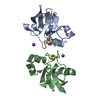
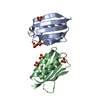
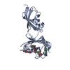
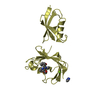
- PDB-1j75: Crystal Structure of the DNA-Binding Domain Zalpha of DLM-1 Bound... -
+ Open data

- Basic information
| Entry | Database: PDB / ID: 1j75 | ||||||
|---|---|---|---|---|---|---|---|
| Title | Crystal Structure of the DNA-Binding Domain Zalpha of DLM-1 Bound to Z-DNA | ||||||
 Components Components |
| ||||||
 Keywords Keywords | IMMUNE SYSTEM/DNA / PROTEIN-Z-DNA COMPLEX / IMMUNE SYSTEM-DNA COMPLEX | ||||||
| Function / homology |  Function and homology information Function and homology informationleft-handed Z-DNA binding / regulation of interleukin-1-mediated signaling pathway / execution phase of necroptosis / double-stranded RNA adenosine deaminase activity / necroptotic signaling pathway / positive regulation of necroptotic process / positive regulation of type I interferon-mediated signaling pathway / pyroptotic inflammatory response / defense response to fungus / activation of innate immune response ...left-handed Z-DNA binding / regulation of interleukin-1-mediated signaling pathway / execution phase of necroptosis / double-stranded RNA adenosine deaminase activity / necroptotic signaling pathway / positive regulation of necroptotic process / positive regulation of type I interferon-mediated signaling pathway / pyroptotic inflammatory response / defense response to fungus / activation of innate immune response / antiviral innate immune response / positive regulation of inflammatory response / double-stranded RNA binding / regulation of inflammatory response / defense response to virus / positive regulation of apoptotic process / apoptotic process / DNA binding / RNA binding / identical protein binding / nucleus / cytoplasm / cytosol Similarity search - Function | ||||||
| Biological species |  | ||||||
| Method |  X-RAY DIFFRACTION / SYNCHROTRON / MOLECULAR REPLACEMENT / Resolution: 1.85 Å X-RAY DIFFRACTION / SYNCHROTRON / MOLECULAR REPLACEMENT / Resolution: 1.85 Å | ||||||
 Authors Authors | Schwartz, T. / Behlke, J. / Lowenhaupt, K. / Heinemann, U. / Rich, A. | ||||||
 Citation Citation | Journal: Nat.Struct.Biol. / Year: 2001 Title: Structure of the DLM-1-Z-DNA complex reveals a conserved family of Z-DNA-binding proteins. Authors: Schwartz, T. / Behlke, J. / Lowenhaupt, K. / Heinemann, U. / Rich, A. | ||||||
| History |
|
- Structure visualization
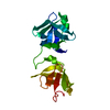
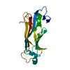
| Structure viewer | Molecule: MolmilJmol/JSmol |
|---|
- Downloads & links
-Download
| PDBx/mmCIF format | 1j75.cif.gz | 32.7 KB | Display | PDBx/mmCIF format |
|---|---|---|---|---|
| PDB format | pdb1j75.ent.gz | 19.1 KB | Display | PDB format |
| PDBx/mmJSON format | 1j75.json.gz | Tree view | PDBx/mmJSON format | |
| Others |  Other downloads Other downloads |
-Validation report
| Arichive directory | https://data.pdbj.org/pub/pdb/validation_reports/j7/1j75ftp://data.pdbj.org/pub/pdb/validation_reports/j7/1j75 | HTTPS FTP |
|---|
-Related structure data
| Related structure data |  1qbjS S: Starting model for refinement |
|---|---|
| Similar structure data |


-Links
 PDBj
PDBj- Assembly
| Deposited unit | 
| ||||||||||||||||||
|---|---|---|---|---|---|---|---|---|---|---|---|---|---|---|---|---|---|---|---|
| 1 | 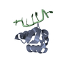
| ||||||||||||||||||
| Unit cell |
| ||||||||||||||||||
| Components on special symmetry positions |
|
-Components
| #1: DNA chain | Mass: 2114.398 Da / Num. of mol.: 1 / Source method: obtained synthetically |
|---|---|
| #2: Protein | Mass: 7328.450 Da / Num. of mol.: 1 / Fragment: N-TERMINAL WINGED-HELIX DOMAIN ZALPHA Source method: isolated from a genetically manipulated source Source: (gene. exp.)  |
| #3: Water | ChemComp-HOH /  Mass: 18.015 Da / Num. of mol.: 102 / Source method: isolated from a natural source / Formula: H2O Mass: 18.015 Da / Num. of mol.: 102 / Source method: isolated from a natural source / Formula: H2O |
-Experimental details
-Experiment
| Experiment | Method: X-RAY DIFFRACTION / Number of used crystals: 2 |
|---|
- Sample preparation
| Crystal | Density Matthews: 2.59 Å3/Da / Density % sol: 52.2 % | ||||||||||||||||||||||||||||||||||||||||||||||||
|---|---|---|---|---|---|---|---|---|---|---|---|---|---|---|---|---|---|---|---|---|---|---|---|---|---|---|---|---|---|---|---|---|---|---|---|---|---|---|---|---|---|---|---|---|---|---|---|---|---|
| Crystal grow | Temperature: 297 K / Method: vapor diffusion, hanging drop / pH: 6 Details: 15% PEG 4000, 0.1M ammonium hydrogen phosphate, 15% ethylene glycol, 0.1M MES pH 6.0, pH 6.00, VAPOR DIFFUSION, HANGING DROP, temperature 297K | ||||||||||||||||||||||||||||||||||||||||||||||||
| Components of the solutions |
| ||||||||||||||||||||||||||||||||||||||||||||||||
| Crystal grow | *PLUS Temperature: 24 ℃ / pH: 7.5 / Method: vapor diffusion | ||||||||||||||||||||||||||||||||||||||||||||||||
| Components of the solutions | *PLUS
|
-Data collection
| Diffraction | Mean temperature: 100 K |
|---|---|
| Diffraction source | Source: SYNCHROTRON / Site: EMBL/DESY, HAMBURG  / Beamline: X11 / Wavelength: 0.9102 / Wavelength: 0.9102 Å / Beamline: X11 / Wavelength: 0.9102 / Wavelength: 0.9102 Å |
| Detector | Type: MARRESEARCH / Detector: CCD / Date: Aug 16, 2000 |
| Radiation | Monochromator: MIRRORS / Protocol: SINGLE WAVELENGTH / Monochromatic (M) / Laue (L): M / Scattering type: x-ray |
| Radiation wavelength | Wavelength: 0.9102 Å / Relative weight: 1 |
| Reflection | Resolution: 1.85→32 Å / Num. all: 7802 / Num. obs: 7802 / % possible obs: 99.8 % / Observed criterion σ(F): 0 / Observed criterion σ(I): 0 / Redundancy: 23.4 % / Biso Wilson estimate: 37.6 Å2 / Rmerge(I) obs: 0.066 / Net I/σ(I): 6.7 |
| Reflection shell | Resolution: 1.85→1.88 Å / Redundancy: 7.9 % / Rmerge(I) obs: 0.613 / Mean I/σ(I) obs: 2.9 / % possible all: 99.5 |
| Reflection | *PLUS Num. measured all: 182894 |
| Reflection shell | *PLUS % possible obs: 99.5 % |
- Processing
| Software |
| ||||||||||||||||||||||||||||
|---|---|---|---|---|---|---|---|---|---|---|---|---|---|---|---|---|---|---|---|---|---|---|---|---|---|---|---|---|---|
| Refinement | Method to determine structure: MOLECULAR REPLACEMENT Starting model: PDB ENTRY 1QBJ Resolution: 1.85→32 Å / Cross valid method: THROUGHOUT / σ(F): 0 / σ(I): 0 / Stereochemistry target values: MAXIMUM LIKELIHOOD / Details: CNS 1.0 and REFMAC5 used in refinement
| ||||||||||||||||||||||||||||
| Solvent computation | Solvent model: BABINET MODEL WITH MASK PARAMETERS FOR MASK CALCULATION VDW PROBE RADIUS : 1.40 ION PROBE RADIUS : 0.80 SHRINKAGE RADIUS : 0.80 | ||||||||||||||||||||||||||||
| Displacement parameters | Biso mean: 21.3 Å2
| ||||||||||||||||||||||||||||
| Refinement step | Cycle: LAST / Resolution: 1.85→32 Å
| ||||||||||||||||||||||||||||
| Refine LS restraints |
| ||||||||||||||||||||||||||||
| LS refinement shell | Resolution: 1.85→1.88 Å
| ||||||||||||||||||||||||||||
| Software | *PLUS Name: CNS / Version: 1 / Classification: refinement | ||||||||||||||||||||||||||||
| Refinement | *PLUS σ(F): 0 / % reflection Rfree: 10.6 % / Rfactor obs: 0.22152 / Rfactor Rfree: 0.24 | ||||||||||||||||||||||||||||
| Solvent computation | *PLUS | ||||||||||||||||||||||||||||
| Displacement parameters | *PLUS Biso mean: 21.3 Å2 | ||||||||||||||||||||||||||||
| LS refinement shell | *PLUS Rfactor Rfree: 0.313 / Rfactor Rwork: 0.279 |
